Review
. 2012 Mar;27 Suppl 2(Suppl 2):65-8.
doi: 10.1111/j.1440-1746.2011.07002.x.

What's new in liver fibrosis? The origin of myofibroblasts in liver fibrosis

Affiliations
Review

What's new in liver fibrosis? The origin of myofibroblasts in liver fibrosis

Keiko Iwaisako et al. J Gastroenterol Hepatol. 2012 Mar.

Abstract

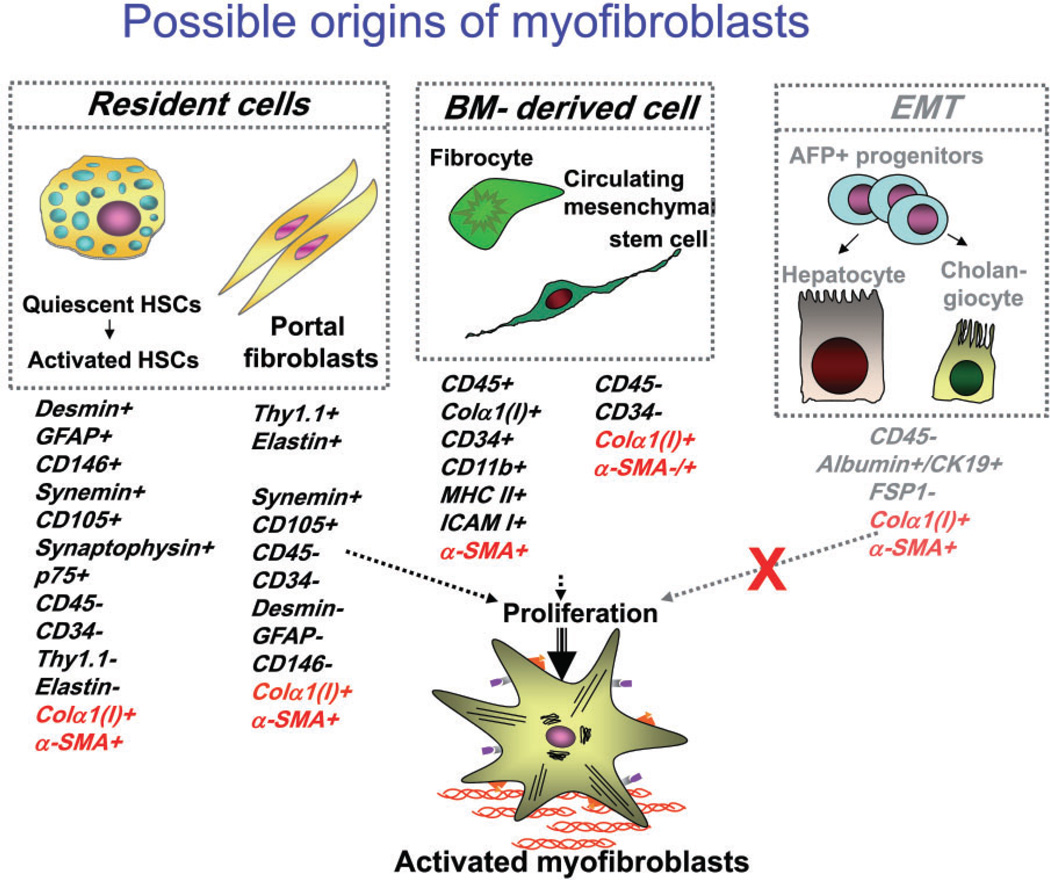
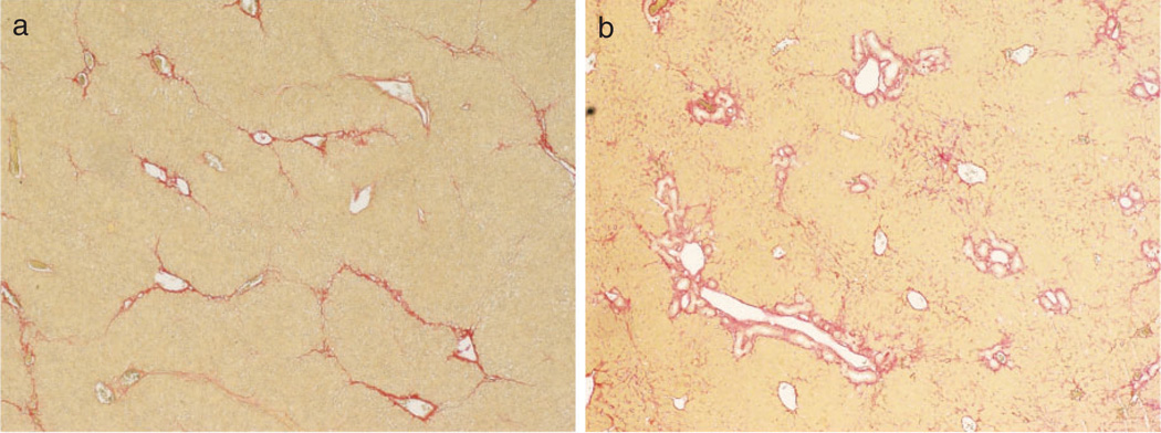
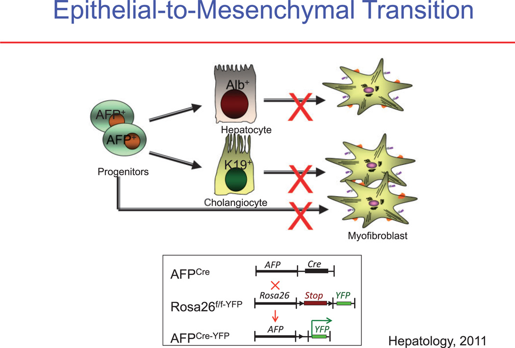
Chronic liver injury of many etiologies produces liver fibrosis and may eventually lead to the formation of cirrhosis. Fibrosis is part of a dynamic process associated with the continuous deposition and resorption of extracellular matrix, mainly fibrillar collagen. Studies of fibrogenesis conducted in many organs including the liver demonstrate that the primary source of the extracellular matrix in fibrosis is the myofibroblast. Hepatic myofibroblasts are not present in the normal liver but transdifferentiate from heterogeneous cell populations in response to a variety of fibrogenic stimuli. Debate still exists regarding the origin of hepatic myofibroblasts. It is considered that hepatic stellate cells and portal fibroblasts have fibrogenic potential and are the major origin of hepatic myofibroblasts. Depending on the primary site of injury the fibrosis may be present in the hepatic parenchyma as seen in chronic hepatitis or may be restricted to the portal areas as in most biliary diseases. It is suggested that hepatic injury of different etiology triggers the transdifferentiation to myofibroblasts from distinct cell populations. Here we discuss the origin and fate of myofibroblast in liver fibrosis.


Conflict of interest statement

Conflict of Interests: There are no conflicts of interests.

Figures

Figure 1
Figure 1
The proposed sources of hepatic myofibroblasts: resident cells (hepatic stellate cells and portal fibroblasts); bone marrow (BM)-derived mesenchymal cells, and cells originated by epithelial-to-mesenchymal transition. Modified from Bataller et al.
Figure 2
Figure 2
Liver fibrosis induced by hepatotoxic injury or cholestatic injury in mice. Liver sections were assessed by Sirius red staining. (a) Hepatotoxic injury model; the fibrotic tissue is initially located in pericentral and perisinusoidal areas. In advance, collagen bands to bridging fibrosis to frank cirrhosis occurs. (b) Cholestatic injury model; the fibrotic tissue is initially located around portal tracts.
Figure 3
Figure 3
Analysis of epithelial-to-mesenchymal transition (EMT). (a) It is proposed that in EMT the myofibroblasts in liver fibrosis originate from hepatic epithelial cells, consisting of hepatocytes (albumin+ [Alb+] cells), cholangiocytes (cytokeratin-19+ [K19+] cells), or progenitor cells (AFP+ cells). (b) Determining the origin of myofibroblasts using cell fate mapping. If a cell expressed α-fetoprotein (AFP), it will be irreversibly genetically labeled. AFP-driven Cre labeled epithelial progenitor cells, cholangiocytes, and hepatocytes, but failed to label any hepatic stellate cells (HSCs) or myofibroblasts. YFP, yellow fluorescent protein.

References

    1. Bataller R, Brenner DA. Liver fibrosis. J. Clin. Invest. 2005;115:209–218. - PMC - PubMed
    1. Iredale JP. Models of liver fibrosis: exploring the dynamic nature of inflammation and repair in a solid organ. J. Clin. Invest. 2007;117:539–548. - PMC - PubMed
    1. Schnabl B, Scholten D, Brenner DA. What is the potential role of antifibrotic agents for the treatment of liver disease? Nat. Clin. Pract. Gastroenterol. Hepatol. 2008;5:496–497. - PubMed
    1. Friedman SL, Bansal MB. Reversal of hepatic fibrosis—fact or fantasy? Hepatology. 2006;43:S82–S88. - PubMed
    1. Kisseleva T, Brenner DA. Fibrogenesis of parenchymal organs. Proc. Am. Thorac. Soc. 2008;5:338–342. - PMC - PubMed

Publication types

Substances