Review
. 2011;31(6):447-58.
doi: 10.1615/critrevimmunol.v31.i6.10.

Transcription factor network regulating CD(+)CD8(+) thymocyte survival

Affiliations
Review

Transcription factor network regulating CD(+)CD8(+) thymocyte survival

Ruiqing Wang et al. Crit Rev Immunol. 2011.

Abstract

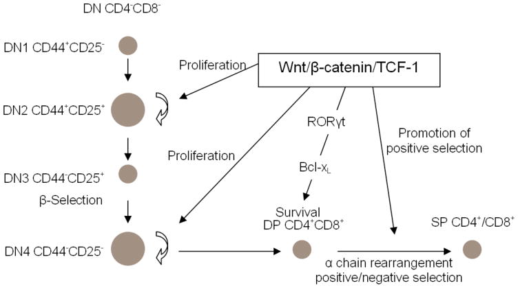
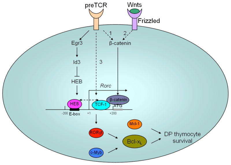
More than 80% of thymocytes are CD4(+)CD8(+) double positive (DP) cells subject to positive/ negative selection. The lifespan of DP thymocytes is critical in shaping the peripheral T-cell repertoire essential for mounting immune responses against foreign, but not self, antigens. During T-cell maturation, if the first round of T-cell receptor (TCR) α chain rearrangement fails to generate a productive T-cell receptor, DP cells start another round of α chain rearrangement until positive selection or cell death intervenes. Thus, the lifespan of DP cells determines how many rounds of α chain rearrangement can be carried out, and influences the likelihood of completing positive selection. The antiapoptotic protein Bcl-x(L) is the ultimate effector regulating DP cell survival, and several transcription factors critical for T-cell development, such as TCF-1, E proteins, c-Myb, and RORγt, regulate DP survival via a Bcl-x(L)-dependent pathway. However, the relationship between these transcription factors in this process is largely unclear. Recent results are revealing an interactive network among these critical factors during regulation of DP thymocyte survival. This review will discuss how these transcription factors potentially work together to control DP thymocyte survival that is critical for successful completion of T-cell development.


Figures

Figure 1
Figure 1
Wnt/β-catenin/TCF-1 regulates thymocyte development at multiple stages. At the DN stage, particularly for DN2 and DN4, β-catenin/TCF-1 promotes thymocyte proliferation to expand the thymocyte pool extensively in order to prepare for β-selection and positive/negative selection. DP thymocyte survival requires β-catenin/TCF-1, which acts upstream of RORγt in the upregulation of Bcl-xL. β-catenin/TCF-1 also promotes positive selection.
Figure 2
Figure 2
Model for possible mechanisms of regulation of DP thymocytes by multiple factors. Activation of RORγt transcription requires cooperation of the transcription factors TCF-1 and HEB. There are three possible pathways responsible for activation of TCF-1. (1) preTCR signals might activate β-catenin, which translocates into the nucleus and switches TCF-1 from a repressor to an activator, eventually leading to induction of RORγt transcription. (2) Activation of β-catenin could also result from the binding of Wnt ligands to the Frizzled receptor in the canonical Wnt signaling pathway. (3) preTCR signals might induce TCF-1 transcription independently of β-catenin. These three possible pathways may be non-exclusive, and could have synergy that leads to maximal activation of TCF-1 at the DN4 and DP stages. Compared to TCF-1, which upon activation functions to stimulate RORγt transcription, HEB seems to mediate fine-tuning of RORγt transcription. At the DN to DP transition, preTCR signals upregulate Egr3 transiently. Egr3-induced Id3 sequesters E proteins such as HEB from binding to the RORγt promoter. Because both HEB and TCF-1 are required for substantial induction of RORγt, the absence of HEB leads to reduction of RORγt expression. When thymocytes differentiate to the DP stage, Egr3 subsides and Id3 is removed, which leads to recovery of HEB activity. HEB then binds to the RORγt promoter, together with already activated TCF-1, and leads to stimulation of maximal RORγt transcription. HEB binds to two E-box sites 200 bp upstream of the RORγt transcription starting site, whereas TCF-1 targets the 200 bp fragment between the RORγt transcription starting site and translation starting site, as determined based on our ChIP assay results. Whether HEB and TCF-1 interact with and influence each other’s activity remains unknown. RORγt and c-Myb cooperate in upregulating Bcl-xL, which eventually leads to protection of DP thymocytes from apoptosis. Mcl-1 could also enhance DP thymocyte survival; however, it is not essential and may only partially compensate for lack of Bcl-xL. Dashed lines indicate pathways and interactions that have not yet been proven.

References

    1. Xie H, Huang Z, Wang R, Sun Z. Regulation of thymocyte survival by transcriptional coactivators. Crit Rev Immunol. 2006;26(6):475–86. - PubMed
    1. Rothenberg EV, Moore JE, Yui MA. Launching the T-cell-lineage developmental programme. Nat Rev Immunol. 2008;8(1):9–21. - PMC - PubMed
    1. Goldrath AW, Bevan MJ. Selecting and maintaining a diverse T-cell repertoire. Nature. 1999;402(6759):255–62. - PubMed
    1. Guo J, Hawwari A, Li H, Sun Z, Mahanta SK, Littman DR, Krangel MS, He YW. Regulation of the TCRalpha repertoire by the survival window of CD4(+)CD8(+) thymocytes. Nat Immunol. 2002;3(5):469–76. - PubMed
    1. Xie H, Huang Z, Sadim MS, Sun Z. Stabilized beta-catenin extends thymocyte survival by up-regulating Bcl-xL. J Immunol. 2005;175(12):7981–8. - PubMed

Publication types

MeSH terms

Substances