. 2012 Apr;11(2):345-9.
doi: 10.1111/j.1474-9726.2012.00795.x. Epub 2012 Feb 9.

A senescent cell bystander effect: senescence-induced senescence

Affiliations
Free PMC article

A senescent cell bystander effect: senescence-induced senescence

Glyn Nelson et al. Aging Cell. 2012 Apr.
Free PMC article

Abstract

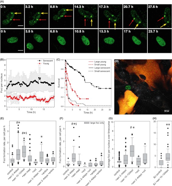
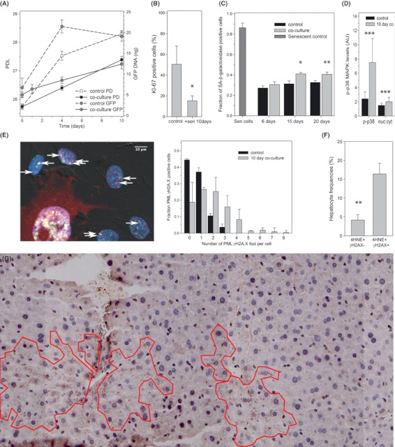
Senescent cells produce and secrete various bioactive molecules including interleukins, growth factors, matrix-degrading enzymes and reactive oxygen species (ROS). Thus, it has been proposed that senescent cells can damage their local environment, and a stimulatory effect on tumour cell growth and invasiveness has been documented. However, it was unknown what effect, if any, senescent cells have on their normal, proliferation-competent counterparts. We show here that senescent cells induce a DNA damage response, characteristic for senescence, in neighbouring cells via gap junction-mediated cell-cell contact and processes involving ROS. Continuous exposure to senescent cells induced cell senescence in intact bystander fibroblasts. Hepatocytes bearing senescence markers clustered together in mice livers. Thus, senescent cells can induce a bystander effect, spreading senescence towards their neighbours in vitro and, possibly, in vivo.


Figures

Fig. 1
Fig. 1
Senescent founder cells induce a senescence-like DDR in bystander cells. (A) Representative images of 53BP1 reporter fluorescence in a senescent (top) or young (bottom) MRC5 cell nucleus with time. Yellow and red arrows indicate two large foci that remain stable over > 27.5 h in the senescent cell. Images are compressed z stacks over 4.5 μm to capture the entire nuclear volume. (B) Mean 53BP1 foci frequencies over time in proliferating (red) and senescent (black) MRC5 cells. Data are mean ± SD of at least 25 nuclei, from three independent experiments. (C) Kaplan–Meier survival curves (censored data) for large (solid lines) and small (dotted lines) 53BP1 foci in proliferating (red) and senescent (black) MRC5 fibroblasts. Foci numbers are 202 (young) and 138 (senescent) from two independent experiments. (D) Representative image of co-cultured senescent founder cells (marked by cytoplasmic RFP staining) and bystander MRC5-AcGFP-53BP1 cells (green nuclear fluorescence). Note nuclear foci in the central bystander cell. (E) 53BP1 foci formation rates in bystander MRC5-AcGFP-53BP1 cells. Co-culture was with no cells without (control) or with either octanol (+oct) or 100 IU SOD and 100 IU catalase (+antiox), with senescent MRC5 cells for 2 days (+sen 2–4 day) or 10 days (+sen 10–12 day) without or with either octanol (+sen 2–4 day +oct) or 100 IU SOD and 100 IU catalase (+sen 2–4 day +antiox) prior to 55 h imaging, or grown in senescent cell conditioned medium for 2 days (+sen med). Box plots indicate median, upper and lower quartiles (boxes), upper and lower centiles (whiskers) and outliers (dots). Significant differences between bystanders and their respective controls (control, +oct or +antiox) are indicated by *, # and $ denote significant differences to antioxidant- or octanol-treated bystanders, respectively (Kruskal–Wallis anova on ranks with Dunn’s post hoc test, P < 0.05). (F) Formation rate of large foci after 2 days co-culture. Data and statistics are as in (D). (G,H) Average foci frequencies per nucleus in bystander MRC5-AcGFP-53BP1 (G) and BJ-AcGFP-53BP1 (H) fibroblasts. Data and statistics are as in (E).
Fig. 2
Fig. 2
Senescent cells induce senescence in surrounding cells. (A) Impact of co-culture with senescent MRC5 cells on growth of MRC5-AcGFP-53BP1 cells measured by either cell counts (square symbols) or GFP-qPCR (circles). Cells were not replated during the experiment, leading to some density-mediated growth inhibition in control cells towards the end of the experiment. However, growth of bystander cells in co-culture was significantly slower (P < 0.01 at 4 and 10 days). (B) Frequencies of Ki67-positive MRC5-AcGFP-53BP1 cells after 10 day culture on their own (control) or 1:1 co-culture with senescent fibroblasts. Data are mean ± SEM, n = 4, P = 0.047. (C) Frequencies of Sen-β-Gal-positive MRC5-AcGFP-53BP1 bystander cells after the indicated time of co-culture with or without (control) senescent MRC5 cells. Senescent inducer cells were removed by blasticidin treatment for 6 days. Increases after 15 and 20 days co-culture are significant (anova with Tukey HSD P = 0.048 and 0.008, respectively). (D) Phospho-p38MAPK (P-p38) levels and nuclear:cytosolic ratios (measured by immunofluorescence) in MRC5-AcGFP-53BP1 after 10 days co-culture. Data are mean ± SD, P < 0.001 (T-test). (E) Co-localization (yellow) of γH2AX foci (green) with PML bodies (red) in bystander BJ-AcGFP-53BP1 cells after 10 day co-culture with senescent cells (marked by red RFP cytoplasmic fluorescence). Left: representative image; right frequency distributions of co-localization-positive cells. Means and distributions are significantly different (P < 0.0000, Mann–Whitney U-test). (F) Percentage of hepatocytes in adult mice liver showing cytoplasmic 4HNE staining together with (4HNE+ γH2AX+) or without (4HNE+ γH2AX−) nuclear DDR. Data are mean ± SE, n = 4 animals. (G) Representative tiled image of mouse liver (9 months old) immunostained for 4-HNE. Areas of clustered 4-HNE-positive cells are outlined. DDR, DNA damage response.

References

    1. Baker DJ, Wijshake T, Tchkonia T, LeBrasseur NK, Childs BG, van de Sluis B, Kirkland JL, van Deursen JM. Clearance of p16Ink4a-positive senescent cells delays ageing-associated disorders. Nature. 2011;479:232–236. - PMC - PubMed
    1. Bavik C, Coleman I, Dean JP, Knudsen B, Plymate S, Nelson PS. The gene expression program of prostate fibroblast senescence modulates neoplastic epithelial cell proliferation through paracrine mechanisms. Cancer Res. 2006;66:794–802. - PubMed
    1. Campisi J, d’Adda di Fagagna F. Cellular senescence: when bad things happen to good cells. Nat. Rev. Mol. Cell Biol. 2007;8:729–740. - PubMed
    1. Chen S, Zhao Y, Zhao G, Han W, Bao L, Yu KN, Wu L. Up-regulation of ROS by mitochondria-dependent bystander signaling contributes to genotoxicity of bystander effects. Mutat. Res. 2009;666:68–73. - PubMed
    1. Coppe JP, Patil CK, Rodier F, Sun Y, Munoz DP, Goldstein J, Nelson PS, Desprez PY, Campisi J. Senescence-associated secretory phenotypes reveal cell-nonautonomous functions of oncogenic RAS and the p53 tumor suppressor. PLoS Biol. 2008;6:2853–2868. - PMC - PubMed

Publication types

Substances