Comparative Study
. 2012 Apr 13;417(5):395-405.
doi: 10.1016/j.jmb.2012.01.026. Epub 2012 Feb 1.

The structures of coiled-coil domains from type III secretion system translocators reveal homology to pore-forming toxins

Affiliations
Comparative Study

The structures of coiled-coil domains from type III secretion system translocators reveal homology to pore-forming toxins

Michael L Barta et al. J Mol Biol. .

Abstract

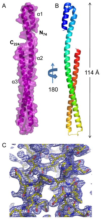
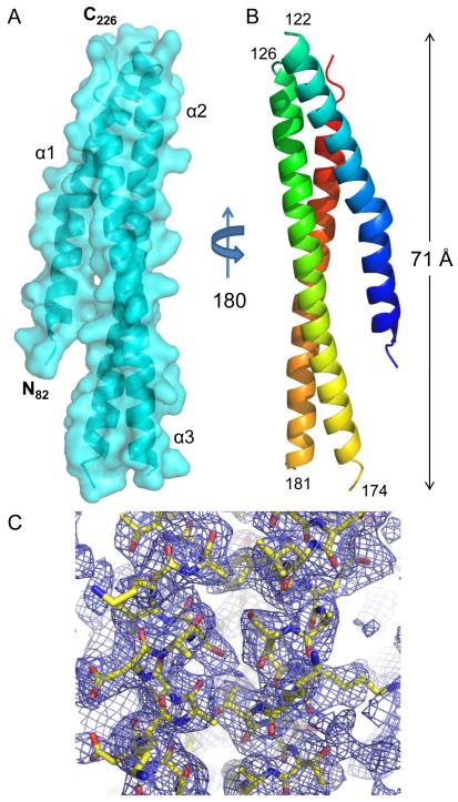
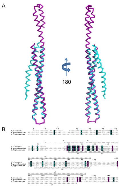
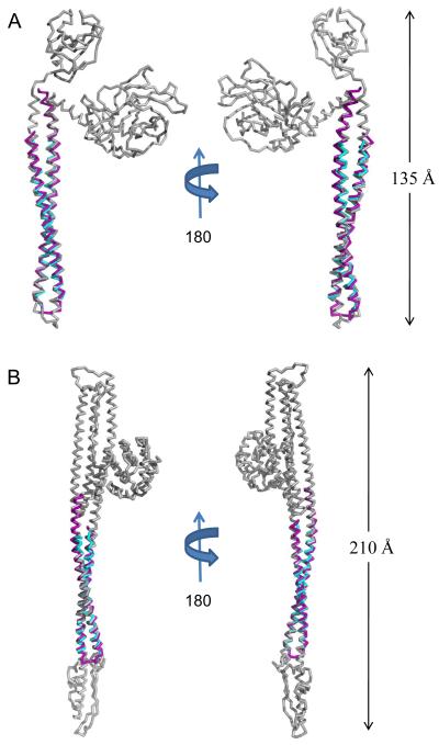
Many pathogenic Gram-negative bacteria utilize type III secretion systems (T3SSs) to alter the normal functions of target cells. Shigella flexneri uses its T3SS to invade human intestinal cells to cause bacillary dysentery (shigellosis) that is responsible for over one million deaths per year. The Shigella type III secretion apparatus is composed of a basal body spanning both bacterial membranes and an exposed oligomeric needle. Host altering effectors are secreted through this energized unidirectional conduit to promote bacterial invasion. The active needle tip complex of S. flexneri is composed of a tip protein, IpaD, and two pore-forming translocators, IpaB and IpaC. While the atomic structure of IpaD has been elucidated and studied, structural data on the hydrophobic translocators from the T3SS family remain elusive. We present here the crystal structures of a protease-stable fragment identified within the N-terminal regions of IpaB from S. flexneri and SipB from Salmonella enterica serovar Typhimurium determined at 2.1 Å and 2.8 Å limiting resolution, respectively. These newly identified domains are composed of extended-length (114 Å in IpaB and 71 Å in SipB) coiled-coil motifs that display a high degree of structural homology to one another despite the fact that they share only 21% sequence identity. Further structural comparisons also reveal substantial similarity to the coiled-coil regions of pore-forming proteins from other Gram-negative pathogens, notably, colicin Ia. This suggests that these mechanistically separate and functionally distinct membrane-targeting proteins may have diverged from a common ancestor during the course of pathogen-specific evolutionary events.


Figures

Fig. 1
Fig. 1. Crystal Structure of IpaB74.224 at 2.1 Å Resolution
A, Crystal structure of S. flexneri IpaB (residues 74-224) shown in cartoon ribbon format surrounded by surface representation (colored purple). Two copies of each polypeptide are found within the asymmetric unit (single copy shown for clarity). B, Crystal structure rotated 180° about the long axis, colored blue (N-terminus) to red (C-terminus). C, Representative model-to-map correlation for IpaB74.224; 2Fo-Fc weighted electron density (contoured at 2.0 σ) is drawn as a blue cage around a region of the coiled-coil. Representations of all structures were generated using PyMol .
Fig. 2
Fig. 2. Crystal Structure of SipB82.226 at 2.8 Å Resolution
A, Crystal structure of S. Typhimurium SipB (residues 82-226) shown in cartoon ribbon format surrounded by surface representation (colored cyan). The electron density map corresponding to solvent exposed loop regions (i.e. residues 123-125 and 175-181) was too weak to model accurately. The start and end of each missing loop region is labeled in panel B. Four copies of SipB are found within the asymmetric unit (single copy shown for clarity). B, Crystal structure rotated 180° about the long axis; colored blue (N-terminus) to red (C-terminus). C, Representative model-to-map correlation for SipB82.226; 2Fo-Fc weighted electron density (contoured at 1.5 σ) is drawn as a blue cage around a region of the coiled-coil. Representations of all structures were generated using PyMol .
Fig. 3
Fig. 3. Structural Superposition of Translocator Coiled-coils
A, Ribbon diagram of a structural alignment of the coiled-coils from IpaB (residues 120-224, purple) and SipB (residues 126-226, cyan) with an RMSD of 1.42 Å over 93/94 Cα atoms within 5.0 Å, rotated 180° about the long axis. Although the overall topology of both structures is similar, there are differences within the N-terminal region spanning the first helix (α1) and turn as well as the length of the second helix (α2). Such differences within the N-terminus of the structures reported here could be reflective of the apparent instability of the chaperone binding domains (CBD) in the absence of their cognate chaperones. B, Limited structure-based sequence alignment of type III secretion first translocators (residues 1-240) colored according to residue conservation (cyan=absolute and purple=similar) as judged by the BLOSUM62 matrix. Alignment was generated using ClustalW and rendered with ESPRIPT. Numbers above the sequences correspond to S. flexneri IpaB. Secondary structure elements of IpaB and SipB are shown above and below the alignment, respectively. Representations of all structures were generated using PyMol . Three-dimensional structures were superimposed using the Local-Global Alignment method (LGA) . Sequence alignments were carried out using CLUSTALW and aligned with secondary structure elements using ESPRIPT .
Fig. 4
Fig. 4. Translocator Coiled-coils Share Structural Homology with Pore-forming Proteins
A, Ribbon diagram of IpaB (residues 104-224, purple) and SipB (residues 126-226, cyan) superimposed upon E. coli Colicin E3 (colored grey; PDB code= 1JCH) with an RMSD of 2.03 Å over 118/121 Cα atoms for IpaB and an RMSD of 1.19 Å over 92/94 Cα atoms for SipB. Crystal structures are rotated 180° about their long axis. B, Ribbon diagram of IpaB (residues 104-224, purple) and SipB (residues 126-226, cyan) superimposed over E. coli Colicin Ia (colored grey; PDB code= 1CII) with an RMSD of 1.73 Å over 121/121 Cα atoms for IpaB and an RMSD of 1.21 Å over 93/94 Cα atoms for SipB. Crystal structures are rotated 180° about their long axis. The DALI server was used to query available structures within the PDB for structural homology . Representations of all structures were generated using PyMol . Three-dimensional structures were superimposed using the Local-Global Alignment method (LGA) .

Similar articles

Cited by

References

    1. Diarrheal Diseases: Shigellosis. Vol. 2009. World Health Organization; 2009. http://www.who.int/vaccine_research/diseases/diarrhoeal/en/index6.html.
    1. Mueller C, Broz P, Cornelis G. The type III secretion system tip complex and translocon. Mol Microbiol. 2008;68:1085–95. - PubMed
    1. Cornelis GR. The type III secretion injectisome, a complex nanomachine for intracellular ‘toxin’ delivery. Biol Chem. 2010;391:745–51. - PubMed
    1. Blocker A, Jouihri N, Larquet E, Gounon P, Ebel F, Parsot C, Sansonetti P, Allaoui A. Structure and composition of the Shigella flexneri “needle complex”, a part of its type III secreton. Mol Microbiol. 2001;39:652–63. - PubMed
    1. Darboe N, Kenjale R, Picking WL, Picking WD, Middaugh CR. Physical characterization of MxiH and PrgI, the needle component of the type III secretion apparatus from Shigella and Salmonella. Protein Sci. 2006;15:543–52. - PMC - PubMed

Publication types

LinkOut - more resources