. 2012 Feb 9;14(1):R27.
doi: 10.1186/bcr3112.

The growth response to androgen receptor signaling in ERα-negative human breast cells is dependent on p21 and mediated by MAPK activation

Affiliations

The growth response to androgen receptor signaling in ERα-negative human breast cells is dependent on p21 and mediated by MAPK activation

Joseph P Garay et al. Breast Cancer Res. .

Abstract

Introduction: Although a high frequency of androgen receptor (AR) expression in human breast cancers has been described, exploiting this knowledge for therapy has been challenging. This is in part because androgens can either inhibit or stimulate cell proliferation in pre-clinical models of breast cancer. In addition, many breast cancers co-express other steroid hormone receptors that can affect AR signaling, further obfuscating the effects of androgens on breast cancer cells.

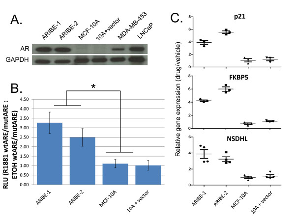
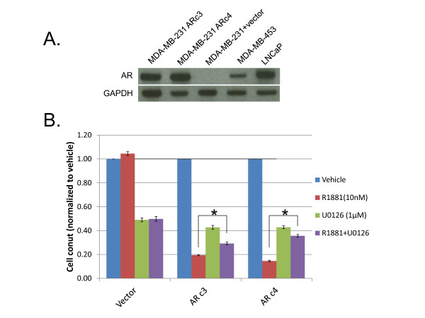
Methods: To create better-defined models of AR signaling in human breast epithelial cells, we took estrogen receptor (ER)-α-negative and progesterone receptor (PR)-negative human breast epithelial cell lines, both cancerous and non-cancerous, and engineered them to express AR, thus allowing the unambiguous study of AR signaling. We cloned a full-length cDNA of human AR, and expressed this transgene in MCF-10A non-tumorigenic human breast epithelial cells and MDA-MB-231 human breast-cancer cells. We characterized the responses to AR ligand binding using various assays, and used isogenic MCF-10A p21 knock-out cell lines expressing AR to demonstrate the requirement for p21 in mediating the proliferative responses to AR signaling in human breast epithelial cells.

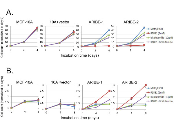
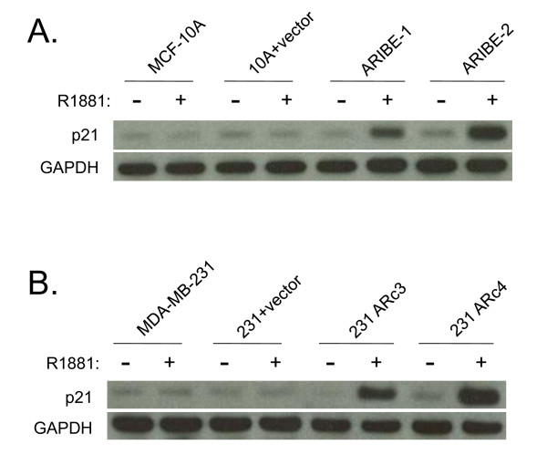
Results: We found that hyperactivation of the mitogen-activated protein kinase (MAPK) pathway from both AR and epidermal growth factor receptor (EGFR) signaling resulted in a growth-inhibitory response, whereas MAPK signaling from either AR or EGFR activation resulted in cellular proliferation. Additionally, p21 gene knock-out studies confirmed that AR signaling/activation of the MAPK pathway is dependent on p21.

Conclusions: These studies present a new model for the analysis of AR signaling in human breast epithelial cells lacking ERα/PR expression, providing an experimental system without the potential confounding effects of ERα/PR crosstalk. Using this system, we provide a mechanistic explanation for previous observations ascribing a dual role for AR signaling in human breast cancer cells. As previous reports have shown that approximately 40% of breast cancers can lack p21 expression, our data also identify potential new caveats for exploiting AR as a target for breast cancer therapy.


Figures

Figure 1
Figure 1
Expression of androgen receptor (AR) in human breast epithelial cells. (A) MCF-10A cells were stably transfected with an AR cDNA, and lysates from single-cell clones were probed for AR expression by western blotting. As a negative control, MCF-10A cells were also transfected with empty vector (10A plus vector) and isolated as single-cell clones. MDA-MB-453 and LNCaP cell lysates served as positive controls for AR expression. All lysates were also probed with GAPDH antibody as a loading control. (B) Androgen Receptor In Breast Epithelium (ARIBE) cells and control cells were transfected with luciferase reporter plasmids containing consensus DNA binding sites and mutated controls for AR as described in Methods. At the time of transfection, cells were treated with 1 nmol/l R1881 or vehicle (ethanol), and analyzed 48 hours later. Firefly luciferase measurements were normalized to Renilla luciferase measurements in all samples. The relative luciferase units (RLU) ratio was calculated as the luciferase expression comparing wild-type ARE/mutant ARE in R1881 versus vehicle-treated control cells (R1881 wtARE/mutARE: ETOH wtARE/mutARE). Error bars represent the standard deviation of three independent experiments. Luciferase expression in each ARIBE clone compared with control cell lines was significant by one-way analysis of variance (ANOVA) (P < 0.05).(C) cDNA was made from RNA of cells treated with 1 nmo/l R1881 or vehicle for 48 hours. Quantitative real-time PCR using SYBR Green was performed on triplicate samples of each cell line using intron-spanning primers for each of three androgen-response genes. All cycle threshold numbers were normalized to a control gene, TATA binding protein (TBP). Ratio is expression in cells treated with drug versus vehicle. Error bars represent the standard deviation of four independent experiments. In both ARIBE lines, induction of all genes after drug treatment was significant by one-way ANOVA compared with control cell lines (P < 0.001).
Figure 2
Figure 2
Activation of the mitogen-activated protein kinase (MAPK) pathway alters the growth response of Androgen Receptor In Breast Epithelium (ARIBE) cells to androgen receptor (AR) ligand. (A) Control cells (MCF-10A and 10A plus vector) and two ARIBE clones were cultured in normal propagation media (with 20 ng/ml epidermal growth factor (EGF)) and treated with vehicle, 10 μmol/l bicalutamide, 1 nmol/l R1881, or a combination of R1881 and bicalutamide. Cells were counted after either 2 or 4 days of treatment, and normalized to values of cells counted on the day of drug addition (day 0). Error bars represent the standard deviation of the mean of three independent cell counts. The growth difference between cells treated with R1881 and R1881 plus bicalutamide was significant by Student's two-tailed t-test (P < 0.001). (B) Cells were cultured under normal propagation conditions except for the absence of EGF from the medium. Cells were treated with vehicle or drugs as above. Cells were counted after either 4 or 8 days of treatment, and normalized to values of cells counted on the day of drug addition (day 0). Error bars represent the standard deviation of the mean of three independent cell counts. The growth difference between cells treated with R1881 and R1881 plus bicalutamide was significant by Student's two-tailed t-test (P < 0.01).
Figure 3
Figure 3
Androgen Receptor In Breast Epithelium (ARIBE) cells stimulate mitogen-activated protein kinase (MAPK) signaling upon androgen receptor (AR) ligand binding. Control cells (MCF-10A and 10A plus vector) and ARIBE cells were cultured under normal propagation conditions in the presence or absence of 20 ng/ml epidermal growth factor (EGF), treated with either vehicle or 1 nmol/l R1881 for 48 hours, and then used to make lysates for western blotting as described in Methods. Blots were probed for phosphorylated extracellular signal-regulated kinase (ERK) (Thr-202/Tyr-204) and total ERK. GAPDH antibody was used as loading control.
Figure 4
Figure 4
MDA-MB-231 cells transfected with androgen receptor (AR) show growth inhibition when treated with androgen. (A) The breast cancer cell line MDA-MB-231 was transfected with an AR cDNA, and lysates from single-cell clones were analyzed for AR protein expression. Two representative clones are shown. MDA-MB-453 and LNCaP lysates served as positive controls for AR expression. Lysates were also probed with GAPDH as a loading control.(B) Two MDA-MB-231 clones transfected with AR (ARc3 and ARc4) and control cells (Vector) were treated with vehicle, 1 μmol/l of the MEK inhibitor U0126, 1 nmol/l of R1881, or a combination of R1881 and U0126. Cells were counted after 4 days of drug treatment, and normalized to counts of vehicle-treated control cells. Error bars represent the standard deviation of the mean of three independent counts. The growth difference between cells treated with R1881 and R1881 plus U0126 was significant by Student's two-tailed t-test (*P < 0.005).
Figure 5
Figure 5
Upregulation of p21 expression upon androgen receptor (AR) ligand binding in the presence of mitogen-activated protein kinase (MAPK)signaling. (A) Control cells (MCF-10A and 10A plus vector) and ARIBE cells were cultured in normal propagation media (with 20 ng/ml EGF) and treated with either vehicle or 1 nmol/l R1881 for 24 hours. Whole-cell lysates were probed for expression of p21 and GAPDH for a loading control. (B) Control cells (MDA-MB-231 and 231 plus vector) and MDA-MB-231 cells expressing AR (ARc3 and ARc4) were cultured in the presence EGF and treated with either vehicle or 1 nmol/l R1881 for 24 hours. Whole-cell lysates were probed for expression of p21 and GAPDH was used as loading control.
Figure 6
Figure 6
p21 knock-down via siRNA abrogates growth inhibition of Androgen Receptor In Breast Epithelium (ARIBE) cells. (A) ARIBE cells were transfected with p21 siRNA constructs, control siRNA, or no siRNA, and incubated for 24 hours. After incubation, whole-cell lysates were probed for expression of p21, and GAPDH was used as a loading control. (B) ARIBE cells transfected for 24 hours with p21 or control siRNA were subsequently treated with vehicle or 1 nmol/l R1881 for 4 days, and cells were counted. The ratio of growth between vehicle and R1881-treated cells was calculated and normalized to untransfected (no siRNA) values to determine the fold increase in growth of siRNA-transfected cells. Error bars represent the standard deviation of the mean of three independent counts. The growth difference between cells transfected with p21 siRNA constructs and mock siRNA was significant by one-way analysis of variance (*P < 0.05).
Figure 7
Figure 7
p21 mediates growth effects of androgen receptor (AR) ligand in MCF-10A cells. (A) MCF-10A p21-/- cells were stably transfected with an AR cDNA, and lysates from single-cell clones were probed for AR expression by western blotting. As a negative control, MCF-10A p21-/- cells were also transfected with empty vector (p21-/- plus vector) and isolated as single-cell clones. Expression of AR was compared with ARIBE cells, which have wild-type p21. MDA-MB-453 and LNCaP cell lysates served as positive controls for AR expression. All lysates were also probed with GAPDH antibody as a loading control. (B) The p21-/- (left) and p21 wild-type (right) cells were cultured in normal propagation media with 20 ng/ml epidermal growth factor (EGF), and treated with 1 nmol/l R1881. Cells were counted after 4 days of treatment, and normalized to values of cells counted on the day of drug addition (day 0). Error bars represent the standard deviation of the mean of three independent cell counts. The growth difference between p21-/- AR-expressing cells and p21-wild-type AR-expressing cells was significant by one-way analysis of variance (ANOVA) (*P < 0.001). (C) Cells were cultured under normal propagation conditions except for the absence of EGF from the medium. Cells were treated with vehicle or drugs as described above. Cells were counted after 8 days of treatment, and normalized to values of cells counted on the day of drug addition (day 0). Error bars represent the standard deviation of the mean of three independent cell counts. The growth difference between p21-/- AR-expressing cells and p21-wild-type AR-expressing cells was significant by one-way ANOVA (*P < 0.01).
Figure 8
Figure 8
p21-/- cells do not stimulate mitogen-activated protein kinase (MAPK) signaling upon androgen receptor (AR) ligand binding. Control cells (MCF-10A p21-/- and p21-/- plus vector) and AR-expressing cells (p21-/- AR-1 and p21-/- AR-4) were cultured under normal propagation conditions in the presence or absence of 20 ng/ml epidermal growth factor (EGF), and treated with either vehicle or 1 nmol/l R1881 for 48 hours. Western blotting was performed on whole-cell lysates. Blots were probed for phosphorylated extracellular signal-regulated kinase (ERK) (Thr-202/Tyr-204) and total ERK. GAPDH antibody was used as a loading control.

Similar articles

Cited by

References

    1. van de Velde CJ, Verma S, van Nes JG, Masterman C, Pritchard KI. Switching from tamoxifen to aromatase inhibitors for adjuvant endocrine therapy in postmenopausal patients with early breast cancer. Cancer Treat Rev. 2009. - PubMed
    1. Cauley JA, Lucas FL, Kuller LH, Stone K, Browner W, Cummings SR. Elevated serum estradiol and testosterone concentrations are associated with a high risk for breast cancer. Study of Osteoporotic Fractures Research Group. Ann Intern Med. 1999;130:270–277. - PubMed
    1. Hankinson SE, Willett WC, Manson JE, Colditz GA, Hunter DJ, Spiegelman D, Barbieri RL, Speizer FE. Plasma sex steroid hormone levels and risk of breast cancer in postmenopausal women. J Natl Cancer Inst. 1998;90:1292–1299. doi: 10.1093/jnci/90.17.1292. - DOI - PubMed
    1. Dimitrakakis C, Bondy C. Androgens and the breast. Breast Cancer Res. 2009;11:212. doi: 10.1186/bcr2413. - DOI - PMC - PubMed
    1. Lea OA, Kvinnsland S, Thorsen T. Improved measurement of androgen receptors in human breast cancer. Cancer Res. 1989;49:7162–7167. - PubMed

Publication types

MeSH terms