. 2012 Apr;180(4):1386-93.
doi: 10.1016/j.ajpath.2011.12.028. Epub 2012 Feb 7.

Ack1 tyrosine kinase activation correlates with pancreatic cancer progression

Affiliations

Ack1 tyrosine kinase activation correlates with pancreatic cancer progression

Kiran Mahajan et al. Am J Pathol. 2012 Apr.

Abstract

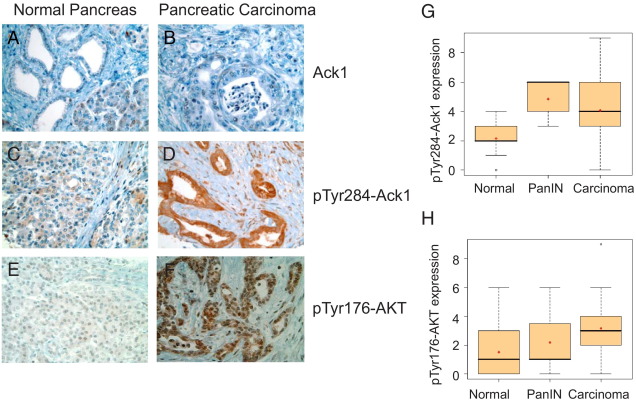
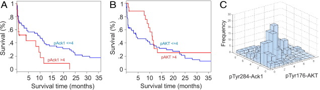
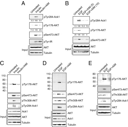
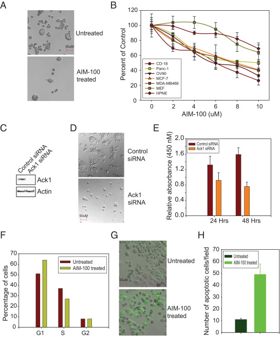
Pancreatic cancer is a significant cause of cancer mortality worldwide as the disease has advanced significantly in patients before symptoms are evident. The signal transduction pathways that promote this rapid progression are not well understood. Ack1 or TNK2, an ubiquitously expressed oncogenic non-receptor tyrosine kinase, integrates signals from ligand-activated receptor tyrosine kinases to modulate intracellular signaling cascades. In the present study, we investigated the Ack1 activation profile in a pancreatic cancer tumor microarray, and observed that expression levels of activated Ack1 and pTyr284-Ack1 positively correlated with the severity of disease progression and inversely correlated with the survival of patients with pancreatic cancer. To explore the mechanisms by which Ack1 promotes tumor progression, we investigated the role of AKT/PKB, an oncogene and Ack1-interacting protein. Ack1 activates AKT directly in pancreatic and other cancer cell lines by phosphorylating AKT at Tyr176 to promote cell survival. In addition, the Ack1 inhibitor AIM-100 not only inhibited Ack1 activation but also suppressed AKT tyrosine phosphorylation, leading to cell cycle arrest in the G1 phase. This effect resulted in a significant decrease in the proliferation of pancreatic cancer cells and induction of apoptosis. Collectively, our data indicate that activated Ack1 could be a prognostic marker for ascertaining early or advanced pancreatic cancer. Thus, Ack1 inhibitors hold promise for therapeutic intervention to inhibit pancreatic tumor growth.


Figures

Figure 1
Figure 1
pTyr284-Ack1 and pTyr176-AKT expressions correlate with disease progression in pancreatic cancer. Shown are TMA sections representing normal and pancreatic cancer stages stained with Ack1 (A and B), pTyr284-Ack1 (C and D) and pTyr176-AKT antibodies (E and F). G and H: Significant increase in pTyr284-Ack1 (G) and pTyr176-AKT (H) expression was seen in PanIN and pancreatic carcinoma as compared with normal pancreatic tissue samples. Spearman's correlation analysis showed that expression levels of both pTyr284-Ack1 and pTyr176-AKT increased significantly from normal to carcinoma (P < 0.0001 for each protein). Boxplots were used to summarize the intensity distribution at each stage. The boxplots have boxes with lines at the lower quartile (25%), median (50%), and upper quartile (75%) values, whereas the red cross within the circle marks the mean value. Whiskers extend from each end of the box to the most extreme values within 1.5 times the interquartile range from the ends of the box. Data with values beyond the ends of the whiskers, indicated with black open circles, are potential outliers.
Figure 2
Figure 2
pTyr284-Ack1 expression correlated negatively with survival in patients with pancreatic cancer. A: Kaplan-Meier analysis shows that patients with pancreatic cancer have higher pTyr284-Ack1 expression levels (with staining intensity >4) and significantly worse overall survival outcome when compared with those with lower expression levels (with staining intensity ≤4) (log-rank test, P = 0.02). B: Kaplan-Meier analysis in patients with pancreatic cancer shows that no significant association was observed between pTyr176-AKT levels and overall survival (log-rank test, P = 0.16). C: Expression of pTyr284-Ack1 was also significantly correlated with that of pTyr176-AKT in pancreatic cancer (Spearman's rank correlation coefficient, ρ = 0.40, P < 0.0001).
Figure 3
Figure 3
AIM-100 inhibited pTyr284-Ack1 and pTyr176-AKT expression in insulin-treated cells. A: Serum-depleted CD18 cells were untreated or were treated with 0.8 μg/mL insulin for 30 minutes and with 10 μmol/L AIM-100 overnight, and lysates were immunoblotted using pTyr284-Ack1, pSer473-AKT, pTyr-IR, panAKT, and tubulin antibodies. The lysates were also immunoprecipitated using pTyr176-AKT antibodies, followed by immunoblotting using AKT antibodies. B: Serum-depleted Panc-1 cells were untreated or were treated with 10 ng/mL EGF for 10 minutes and with 5 and 10 μmol/L AIM-100 overnight, and lysates were immunoblotted using pTyr284-Ack1, pSer473-AKT, panAKT, and tubulin antibodies. The lysates were also immunoprecipitated using pTyr176-AKT antibodies, followed by immunoblotting using AKT antibodies. C–E: Serum-depleted MCF-7 (C), H292 (D), and A2780-CP (E) cells were untreated or were treated with 0.8 μg/mL insulin for 30 minutes or 10 ng/mL EGF for 10 minutes and with 0.8 μmol/L AIM-100 overnight, and lysates were immunoblotted using pTyr284-Ack1, pSer473-AKT, pThr308-AKT, PanAKT, Ack1, and tubulin antibodies. The lysates were also immunoprecipitated using pTyr176-AKT antibodies, followed by immunoblotting using AKT antibodies (top panels).
Figure 4
Figure 4
AIM-100 inhibited growth of pancreatic cancer cells. A: CD18 cells were untreated or were treated with 10 μmol/L AIM-100 for 48 hours, and cells were photographed at 10× magnification using differential interference contrast imaging. AIM-100 treatment significantly inhibited the growth of cells. B: CD18, Panc-1, HPNE, OV90, MCF-7, MDA-MB-468, and MEF cells were untreated or were treated with 2 to 10 μmol/L AIM-100 for 48 hours, and an MTT assay was performed. The experiment was performed twice with eight replicates; a representative data set is shown. C: CD18 cells were electroporated (Lonza) with control and Ack1 siRNA, and cell lysates were immunoblotted using Ack1 and actin antibodies. D: CD18 cells were transfected with Ack1 or control siRNA, and cells were photographed at 10× magnification using differential interference contrast imaging. E: CD18 cells were transfected with Ack1 or control siRNA, and a WST-1 cell proliferation assay was performed. F: Cell cycle analysis was performed using flow cytometry. Panc-1 cells were untreated or were treated with 6 μmol/L AIM-100 for 48 hours. Cells were stained with propidium iodide, and DNA content was measured. The experiment was performed three times, and a representative data set is shown. G: Panc-1 cells were untreated or were treated with 10 μmol/L AIM-100 for 48 hours. The cell death/caspase reagent (Invitrogen Corp.) was added, and caspase activity was assessed by monitoring green fluorescence using a microscope. H: The number of cells positive for green fluorescence was counted in multiple fields, and the mean number per field is plotted.

References

    1. Greenlee R.T., Murray T., Bolden S., Wingo P.A. Cancer Statistics, 2000. CA Cancer J Clin. 2000;50:7–33. - PubMed
    1. Burris H., 3rd, Rocha-Lima C. New therapeutic directions for advanced pancreatic cancer: targeting the epidermal growth factor and vascular endothelial growth factor pathways. Oncologist. 2008;13:289–298. - PubMed
    1. Xiong H.Q., Abbruzzese J.L. Epidermal growth factor receptor–targeted therapy for pancreatic cancer. Semin Oncol. 2002;29:31–37. - PubMed
    1. Moore M.J., Goldstein D., Hamm J., Figer A., Hecht J.R., Gallinger S., Au H.J., Murawa P., Walde D., Wolff R.A., Campos D., Lim R., Ding K., Clark G., Voskoglou-Nomikos T., Ptasynski M., Parulekar W. Erlotinib plus gemcitabine compared with gemcitabine alone in patients with advanced pancreatic cancer: a phase III trial of the National Cancer Institute of Canada Clinical Trials Group. J Clin Oncol. 2007;25:1960–1966. - PubMed
    1. Manning B.D., Cantley L.C. AKT/PKB signaling: navigating downstream. Cell. 2007;129:1261–1274. - PMC - PubMed

Publication types

MeSH terms