. 2012 Apr;180(4):1535-46.
doi: 10.1016/j.ajpath.2011.12.036. Epub 2012 Feb 7.

Intestinal mast cell levels control severity of oral antigen-induced anaphylaxis in mice

Affiliations

Intestinal mast cell levels control severity of oral antigen-induced anaphylaxis in mice

Richard Ahrens et al. Am J Pathol. 2012 Apr.

Abstract

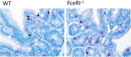
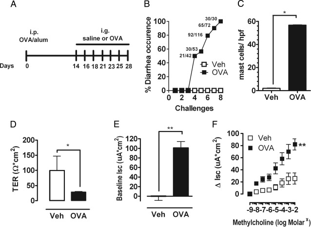
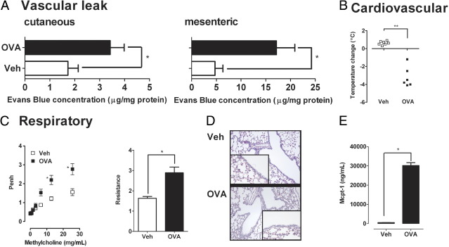
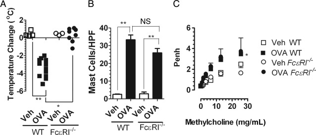
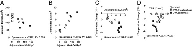
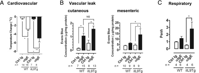
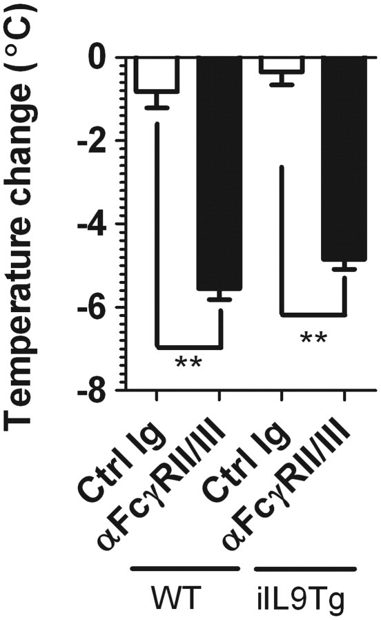
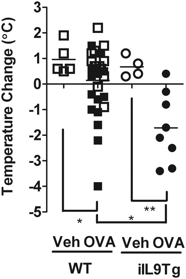
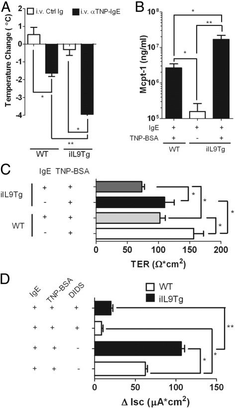
Food-triggered anaphylaxis can encompass a variety of symptoms that affect multiple organ systems and can be life threatening. The molecular distinction between non-life-threatening and life-threatening modes of such anaphylaxis has not yet been delineated. In this study, we sought to identify the specific immune functions that regulate the severity of oral antigen-induced anaphylaxis. We thus developed an experimental mouse model in which repeated oral challenge of ovalbumin-primed mice induced an FcεRI- and IgE-dependent oral antigen-triggered anaphylaxis that involved multiple organ systems. Strikingly, the severity of the systemic symptoms of anaphylaxis (eg, hypothermia) positively correlated with the levels of intestinal mast cells (r = -0.53; P < 0.009). In addition, transgenic mice with both increased intestinal and normal systemic levels of mast cells showed increased severity of both intestinal and extra-intestinal symptoms of IgE-mediated passive as well as oral antigen- and IgE-triggered anaphylaxis. In conclusion, these observations indicate that the density of intestinal mast cells controls the severity of oral antigen-induced anaphylaxis. Thus, an awareness of intestinal mast cell levels in patients with food allergies may aid in determining their susceptibility to life-threatening anaphylaxis and may eventually aid in the treatment of food-triggered anaphylaxis.


Figures

Supplemental Figure S1
Histologic assessment of intestinal mast cells in anaphylactic WT and FceRI−/− mice. Histologic assessment of chloroacetate esterase (CAE)-stained small intestine of OVA-sensitized, i.g. OVA-challenged WT and FceRI−/− mice. Arrows indicate remnants of extracellular mast cell granules (CAE+ granules) in the lamina propria of the small intestine of WT OVA mice, whereas mast cells in FceRI−/− mice remained intact, and granules were confined within the cytoplasm.
Figure 1
Figure 1
Intestinal symptoms in oral antigen-triggered anaphylaxis. A: Oral antigen-triggered anaphylaxis experimental regime. Diarrhea occurrence (B) and mean number of mast cells per high-power field (hpf) (C) in the small intestine of OVA-sensitized, i.g. Veh- or OVA-challenged BALB/c WT mice. The common fractions in panel B represent the number of mice that experienced diarrhea over the number of total mice challenged. TER (D), Isc (E), and β-methylcholine-stimulated (F) changes in Isc (ΔIsc) in jejunum segments of OVA-sensitized, i.g. Veh- or OVA-challenged (seventh challenge) BALB/c WT mice. Values represent mean ± SEM; n = 6 to 10 mice per group. Statistical significance is *P < 0.05 or **P < 0.01.
Figure 2
Figure 2
Systemic manifestations in oral antigen-triggered anaphylaxis. A: Before the seventh OVA challenge, mice received i.v. injection of 2% Evans Blue (200 μL/PBS) and were subsequently challenged with OVA. Sixty minutes after OVA challenge, the mice were sacrificed, and Evans blue concentration was determined in the ear (cutaneous) and intestine (mesenteric). Temperature change from 0 to 60 minutes (B), lung function (C), histology (D), and BALF Mcpt-1 levels (E) after the seventh i.g. Veh or OVA challenge in OVA-sensitized WT mice. C: Airway reactivity to methacholine was measured 30 minutes after the seventh OVA challenge with the use of whole-body plethysmography and invasive technique (flexiVent system). D: Original magnification, ×40. Data in A and C are represented as mean ± SEM; n = 4 to 8 mice per group from triplicate experiments. *P < 0.05; **P < 0.01.
Figure 3
Figure 3
Intestinal and systemic manifestations of food-triggered anaphylaxis is FcεRI/IgE dependent. Temperature change from 0 to 60 minutes and diarrhea (A) and mast cells per high-power field (HPF) in small bowel in WT and FceRI−/− mice (B) after the seventh OVA challenge. Solid symbols indicate mice that developed diarrhea. C: Lung function in OVA-sensitized, i.g. Veh- or OVA-challenged WT and FceRI−/− mice. Airway reactivity to methacholine was measured 30 minutes after the seventh Veh or OVA challenge with the use of whole-body plethysmography. Data in B and C are represented as mean ± SEM; n = 4 to 8 mice per group from duplicate experiments. *P < 0.05; **P < 0.01. NS, not significant.
Figure 4
Figure 4
Correlation between intestinal mast cell levels and intestinal and systemic symptoms of oral antigen-induced anaphylaxis. Spearman's rank correlation coefficient between mean number of mast cells per high-power field (hpf) in the small intestine and small intestine epithelial permeability (TER) (A), or intestinal epithelial chloride secretion (B), and between mean temperature change from 0 to 60 minutes and intestinal epithelial chloride secretion (C), or intestinal epithelial permeability change (D) after the fourth i.g. Veh or OVA challenge in OVA-sensitized WT mice.
Figure 5
Figure 5
Systemic and intestinal manifestations of anti-IgE-treated WT and iIL9Tg mice. Maximum temperature change (A), cutaneous (ear) and mesenteric leak (B), and lung function (C) in BALB/c WT and iIL9Tg mice treated with isotype control (Ig, clone βGL117) or anti-IgE (clone EM95) antibody. B: Before the anti-IgE treatment, mice received i.v. injection of 2% Evans blue (200 μL/PBS) and were subsequently challenged with anti-IgE. C: WT and iIL9Tg mice received an i.v. anti-IgE injection, and airway reactivity to methacholine was measured by whole-body plethysmography. Number of mice are indicated. Data are presented as mean ± SEM *P < 0.05; **P < 0.01. Ctrl Ig, isotype control; NS, not significant.
Figure 6
Figure 6
Anti-FcγRII/III-mediated passive systemic anaphylaxis in WT and iIL9Tg mice. Maximum temperature change of WT and iIL9Tg mice to i.v. injection of anti-FcγRII/III mAb (clone 2.4G2) or isotype control (Ig, clone J1.2) antibody. Data represent mean ± SEM; n = 4 mice per group from duplicate experiments. **P < 0.01 compared with WT control (Ctrl) Ig. Ctrl Ig, isotype control.
Figure 7
Figure 7
Intestinal and systemic manifestations of oral antigen-triggered anaphylaxis in WT and iIL9Tg mice. Temperature change from 0 to 60 minutes and diarrhea in OVA-sensitized, i.g. Veh- or OVA-challenged BALB/c WT and iIL9Tg mice after the seventh OVA challenge. Solid symbols indicate mice that developed diarrhea. *P < 0.05; **P < 0.01.
Figure 8
Figure 8
Passive oral antigen-triggered IgE-mediated anaphylaxis in WT and iIL9Tg mice. Maximum temperature change (A), serum Mcpt-1 levels (B), TER (C), and carbachol-induced change in Isc (ΔIsc) (D) of the small intestine of anti-TNP-IgE- (10 μg) or control Ig- (10 μg) treated WT and iIL9Tg mice orally gavaged with TNP-BSA (50 mg/mL; 500 μL). Values represent mean ± SEM; n = 4 to 8 mice per group. *P < 0.05; **P < 0.01. Ctrl Ig, isotype control.

References

    1. Sampson H.A. Anaphylaxis and emergency treatment. Pediatrics. 2003;111:1601–1608. - PubMed
    1. Ross M.P., Ferguson M., Street D., Klontz K., Schroeder T., Luccioli S. Analysis of food-allergic and anaphylactic events in the national electronic injury surveillance system. J Allergy Clin Immunol. 2008;121:166–171. - PubMed
    1. De Smit V., Cameron P.A., Rainer T.H. Anaphylaxis presentations to an emergency department in Hong Kong: incidence and predictors of biphasic reactions. J Emerg Med. 2005;28:381–388. - PubMed
    1. Brown A.F., McKinnon D., Chu K. Emergency department anaphylaxis: a review of 142 patients in a single year. J Allergy Clin Immunol. 2001;108:861–866. - PubMed
    1. Simons E.R., Chad Z.H., Gold M. Anaphylaxis in children: realtime reporting from a national network. Allergy Clin Immunol Int J World Allergy Org. 2004;(Supplement 1):242.

Publication types

MeSH terms