

A study on the reproducibility of cephalometric landmarks when undertaking a three-dimensional (3D) cephalometric analysis
- PMID: 22322503
- PMCID: PMC3476034
- DOI: 10.4317/medoral.17721
A study on the reproducibility of cephalometric landmarks when undertaking a three-dimensional (3D) cephalometric analysis
Abstract
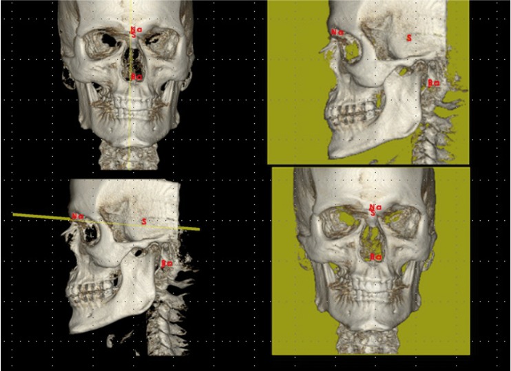
Objectives: Cone Beam Computerized Tomography (CBCT) allows the possibility of modifying some of the diagnostic tools used in orthodontics, such as cephalometry. The first step must be to study the characteristics of these devices in terms of accuracy and reliability of the most commonly used landmarks. The aims were 1- To assess intra and inter-observer reliability in the location of anatomical landmarks belonging to hard tissues of the skull in images taken with a CBCT device, 2- To determine which of those landmarks are more vs. less reliable and 3- To introduce planes of reference so as to create cephalometric analyses appropriated to the 3D reality.
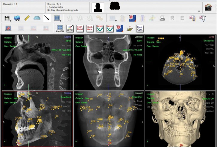
Study design: Fifteen patients who had a CBCT (i-CAT) as a diagnostic register were selected. To assess the reproducibility on landmark location and the differences in the measurements of two observers at different times, 41 landmarks were defined on the three spatial axes (X,Y,Z) and located. 3.690 measurements were taken and, as each determination has 3 coordinates, 11.070 data were processed with SPSS statistical package. To discover the reproducibility of the method on landmark location, an ANOVA was undertaken using two variation factors: time (t1, t2 and t3) and observer (Ob1 and Ob2) for each axis (X, Y and Z) and landmark. The order of the CBCT scans submitted to the observers (Ob1, Ob2) at t1, t2, and t3, were different and randomly allocated. Multiple comparisons were undertaken using the Bonferroni test. The intra- and inter-examiner ICC's were calculated.
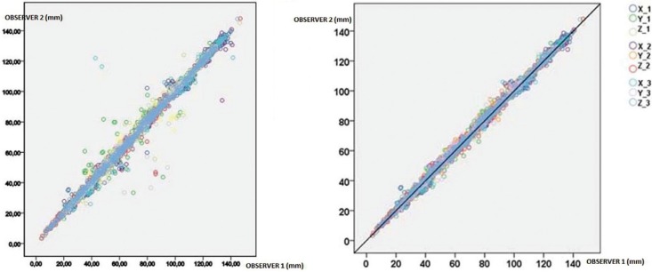
Results: Intra- and interexaminer reliability was high, both being ICC ≥ 0.99, with the best frequency on axis Z.
Conclusions: The most reliable landmarks were: Nasion, Sella, Basion, left Porion, point A, anterior nasal spine, Pogonion, Gnathion, Menton, frontozygomatic sutures, first lower molars and upper and lower incisors. Those with less reliability were the supraorbitals, right zygion and posterior nasal spine.
Figures
References
-
- Mozzo P, Procacci C, Tacconi A, Martini PT, Andreis IA. A new volumetric CT machine for dental imaging based on the cone-beam technique: preliminary results. Eur Radiol. 1998;8:1558–64. - PubMed
-
- Quintero JC, Trosien A, Hatcher D, Kapila S. Craniofacial imaging in orthodontics: Historical perspective, current status, and future developements. Angle Orthod. 1999;69:491–506. - PubMed
-
- Silva MA, Wolf U, Heinicke F, Bumann A, Visser H, Hirsch E. Cone-beam computed tomography for routine orthodontic treatment planning: a radiation dose evaluation. Am J Orthod Dentofacial Orthop. 2008;133:640.e1–5. - PubMed
-
- Chau AC, Fung K. Comparison of radiation dose for implant imaging using conventional spiral tomography, computed tomography, and cone-beam computed tomography. Oral Surg Oral Med Oral Pathol Oral Radiol Endod. 2009;107:559–65. - PubMed
-
- De Vos W, Casselman J, Swennen GR. Cone-beam computerized tomography (CBCT) imaging of the oral and maxillofacial region: a systematic review of the literature. Int J Oral Maxillofac Surg. 2009;38:609–25. - PubMed
MeSH terms
LinkOut - more resources
Full Text Sources
Medical
Miscellaneous
