

Immunohistochemical expression of EGFR in oral leukoplakia: association with clinicopathological features and cellular proliferation
- PMID: 22322523
- PMCID: PMC3482515
- DOI: 10.4317/medoral.17950
Immunohistochemical expression of EGFR in oral leukoplakia: association with clinicopathological features and cellular proliferation
Abstract
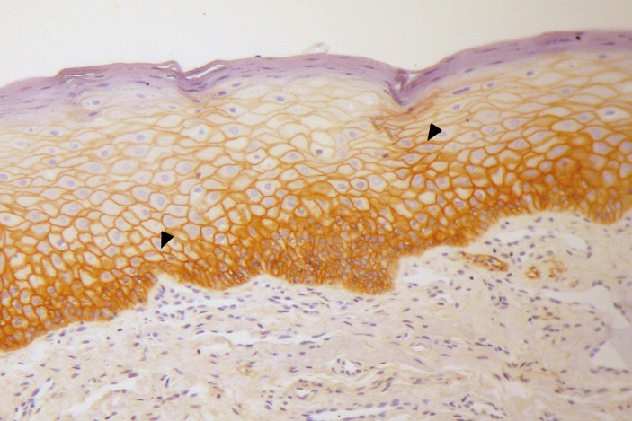
Objectives: To investigate the immunoexpression of epidermal growth factor receptor (EGFR) in a sample of oral leukoplakias (OL) and to determine the receptor' s association with dysplasia, tobacco consumption, lesion site, and proliferation rate. Although EGFR should be overexpressed in some oral leukoplakias, the factors that may interfere with this expression and the influence of this receptor on epithelial proliferation have yet to be investigated.
Study design: Samples of oral leukoplakias (48) and of normal oral epithelium (10) were immunohistologically examined for expression of EGFR. Immunohistochemistry for Ki-67, and p27 were also performed in leukoplakias. EGFR expression was associated with clinical and pathological features.
Results: EGFR was positive in 62.5% of the leukoplakias and 50% of normal oral epithelium. The number of EGFR positive OL located in high-risk sites was significantly higher than EGFR positive OL located in low-risk sites. Most of the p27 negative leukoplakias were EGFR positive, and the p27 index in the parabasal layer was diminished in the presence of dysplasia. Positivity for EGFR was not associated with dysplasia, tobacco exposure, or Ki-67.
Conclusion: EGFR is expressed in leukoplakia regardless of dysplasia, but EGFR positivity should be more frequent in lesions sited in areas of high cancer risk. The association between EGFR and p27 may represent an important mechanism in the control of cellular proliferation and malignant progression of oral epithelium and therefore warrants further investigation.
Figures
Similar articles
-
Expression of p53, epidermal growth factor receptor, c-erbB2 in oral leukoplakias and oral squamous cell carcinomas.J Cancer Res Ther. 2018 Jan-Mar;14(2):388-393. doi: 10.4103/0973-1482.191027. J Cancer Res Ther. 2018. PMID: 29516925
-
Chewing tobacco may act as a risk factor for dysplastic transformation of squamous cells in Oral leukoplakia- A cytochemistry based approach.Pathol Res Pract. 2021 Feb;218:153287. doi: 10.1016/j.prp.2020.153287. Epub 2020 Dec 24. Pathol Res Pract. 2021. PMID: 33454586
-
[Relationship of oral leukoplakia and cancer by immunohistochemical expression of EGF-receptor].Kokubyo Gakkai Zasshi. 1990 Mar;57(1):187-200. doi: 10.5357/koubyou.57.187. Kokubyo Gakkai Zasshi. 1990. PMID: 2370442 Japanese.
-
Markers of proliferation in normal and leukoplakic oral epithelia.Oral Oncol. 2000 Mar;36(2):145-51. doi: 10.1016/s1368-8375(99)00076-7. Oral Oncol. 2000. PMID: 10745166 Review.
-
Unraveling the Keratin Expression in Oral Leukoplakia: A Scoping Review.Int J Mol Sci. 2024 May 21;25(11):5597. doi: 10.3390/ijms25115597. Int J Mol Sci. 2024. PMID: 38891785 Free PMC article.
Cited by
-
Quantitative Immunoexpression of EGFR in Oral Potentially Malignant Disorders: Oral Leukoplakia and Oral Submucous Fibrosis.J Dent Res Dent Clin Dent Prospects. 2015 Summer;9(3):166-74. doi: 10.15171/joddd.2015.031. Epub 2015 Sep 16. J Dent Res Dent Clin Dent Prospects. 2015. PMID: 26697149 Free PMC article.
-
Molecular Analysis as a Guide to Determining the Extent and Pathophysiology of Perilesional Tissues in Oral Epithelial Dysplasias.J Maxillofac Oral Surg. 2020 Sep;19(3):447-455. doi: 10.1007/s12663-019-01303-z. Epub 2019 Nov 11. J Maxillofac Oral Surg. 2020. PMID: 32801543 Free PMC article.
-
Association of Surgical Margins and Pathological Staging with Epidermal Growth Factor Receptor Expression in Oral Squamous Cell Carcinoma- A Prospective Cohort Study.Indian J Otolaryngol Head Neck Surg. 2022 Dec;74(Suppl 3):6001-6006. doi: 10.1007/s12070-021-02629-2. Epub 2021 May 29. Indian J Otolaryngol Head Neck Surg. 2022. PMID: 36742945 Free PMC article.
-
Oral leukoplakia and the long-term risk of upper gastrointestinal cancer deaths in the Linxian dysplasia population.Thorac Cancer. 2020 Oct;11(10):2804-2811. doi: 10.1111/1759-7714.13595. Epub 2020 Aug 18. Thorac Cancer. 2020. PMID: 32808454 Free PMC article.
-
Molecular markers associated with potentially malignant oral lesions (Review).Exp Ther Med. 2021 Aug;22(2):834. doi: 10.3892/etm.2021.10266. Epub 2021 Jun 4. Exp Ther Med. 2021. PMID: 34149880 Free PMC article. Review.
References
-
- Warnakulasuriya S, Johnson NW, van der Waal I. Nomenclature and classification of potentially malignant disorders of the oral mucosa. J Oral Pathol Med. 2007;36:575–80. - PubMed
-
- van der Waal I. Potentially malignant disorders of the oral and oropharyngeal mucosa; terminology, classification and present concepts of management. Oral Oncol. 2009;45:317–23. - PubMed
-
- Warnakulasuriya S, Reibel J, Bouquot J, Dabelsteen E. Oral epithelial dysplasia classification systems: predictive value, utility, weaknesses and scope for improvement. J Oral Pathol Med. 2008;37:127–33. - PubMed
-
- Dietrich T, Reichart PA, Scheifele C. Clinical risk factors of oral leukoplakia in a representative sample of the US population. Oral Oncol. 2004;40:158–63. - PubMed
MeSH terms
Substances
LinkOut - more resources
Full Text Sources
Research Materials
Miscellaneous
