. 2012 Apr;23(7):1157-66.
doi: 10.1091/mbc.E11-09-0772. Epub 2012 Feb 9.

The human mitochondrial ISCA1, ISCA2, and IBA57 proteins are required for [4Fe-4S] protein maturation

Affiliations

The human mitochondrial ISCA1, ISCA2, and IBA57 proteins are required for [4Fe-4S] protein maturation

Alex D Sheftel et al. Mol Biol Cell. 2012 Apr.

Abstract

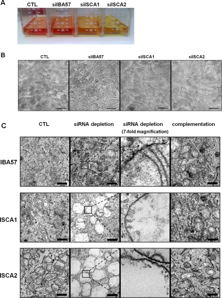
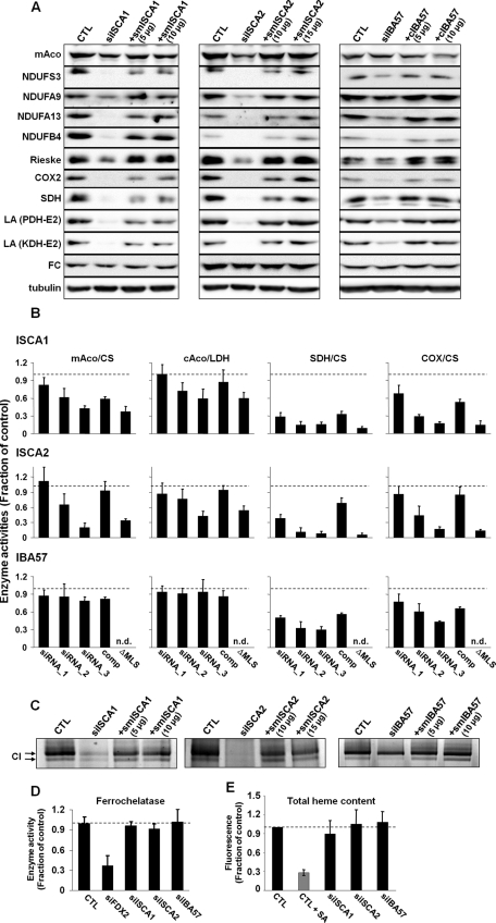
Members of the bacterial and mitochondrial iron-sulfur cluster (ISC) assembly machinery include the so-called A-type ISC proteins, which support the assembly of a subset of Fe/S apoproteins. The human genome encodes two A-type proteins, termed ISCA1 and ISCA2, which are related to Saccharomyces cerevisiae Isa1 and Isa2, respectively. An additional protein, Iba57, physically interacts with Isa1 and Isa2 in yeast. To test the cellular role of human ISCA1, ISCA2, and IBA57, HeLa cells were depleted for any of these proteins by RNA interference technology. Depleted cells contained massively swollen and enlarged mitochondria that were virtually devoid of cristae membranes, demonstrating the importance of these proteins for mitochondrial biogenesis. The activities of mitochondrial [4Fe-4S] proteins, including aconitase, respiratory complex I, and lipoic acid synthase, were diminished following depletion of the three proteins. In contrast, the mitochondrial [2Fe-2S] enzyme ferrochelatase and cellular heme content were unaffected. We further provide evidence against a localization and direct Fe/S protein maturation function of ISCA1 and ISCA2 in the cytosol. Taken together, our data suggest that ISCA1, ISCA2, and IBA57 are specifically involved in the maturation of mitochondrial [4Fe-4S] proteins functioning late in the ISC assembly pathway.


Figures

FIGURE 1:
FIGURE 1:
ISCA1, ISCA2, and IBA57 are localized to mitochondria. (A) HeLa cells were treated with digitonin and centrifuged at 15,000 × g to separate the cell lysate (L) into a membrane fraction containing mitochondria (M) and a cytosolic fraction (C). Localization of ISCA1, ISCA2, and IBA57 was analyzed by immunoblotting using antibodies raised against the respective proteins. Antibodies recognizing the α and β subunits of F1-ATP synthase (F1α/β) and tubulin served to estimate the efficiency of separating mitochondrial and cytosolic proteins, respectively. (B) HeLa cells were cotransfected twice with smISCA1-GFP, smISCA2-GFP, or cIBA57-GFP, together with mitochondria-targeted Discosoma sp. red protein (dsRedMito). Images of living cells were acquired by confocal microscopy.
FIGURE 2:
FIGURE 2:
RNAi depletion of ISCA1, ISCA2, or IBA57 and the effects on the corresponding partner proteins. HeLa cells were transfected with (A) siISCA1, (B) siISCA2, or (C) siIBA57. Cells were harvested after 3 d of growth, and a fraction of the cells was retransfected. This procedure was performed twice. Cell lysates of all three transfection rounds (1–3) and of mock-transfected control cells (CTL) were examined by immunoblotting to compare respective protein levels of ISCA1, ISCA2, and IBA57. Immunostaining against F1α/β and tubulin served as loading controls.
FIGURE 3:
FIGURE 3:
Complementing proteins smISCA1, smISCA2 and cIBA57 functionally localize to mitochondria. HeLa cells were transfected three times as in Figure 2 with specific siRNAs as indicated. Cells were harvested 3 d after the third transfection and fractionated by digitonin treatment as in Figure 1. Cell extracts were analyzed by immunoblotting for the indicated proteins using F1α/β ATP synthase and tubulin as loading controls. Additional samples were cotransfected with the indicated siRNAs plus a vector encoding the corresponding ISC proteins for complementation testing (+smISCA1, +smISCA2, and +cIBA57). In the cases of ISCA1 (A) and ISCA2 (B) these vectors were resistant to RNAi due to silent mutations (sm). Cells were also cotransfected with smISCA1 or smISCA2 vectors lacking the coding information for the mitochondrial localization sequences (ΔMLS). In the case of IBA57 (C) no mutagenesis was necessary to create complementing IBA57 (cIBA57) since the used siRNA bound at the 3′ untranslated region. (D) HeLa cells were transfected with vectors smISCA1ΔMLS and smISCA2ΔMLS, respectively, and 48 h after transfection, a subset of cells was grown in the presence of 5 or 10 μM of the proteasome inhibitor MG132 for 16 h. Harvested cells were lysed, and cell extracts were subjected to immunostaining. preISCA2, putative precursor form of ISCA2.
FIGURE 4:
FIGURE 4:
A decrease in ISCA1, ISCA2, or IBA57 causes severe physiological and morphological changes. HeLa cells were transfected as depicted in Figure 3. (A) Culture media of cells thrice transfected with siISCA1, siISCA2, or siIBA57 or mock-treated control cells (CTL) are shown. The color change of the pH indicator (phenol red) from red toward yellow indicates a drop in pH of the media. (B) After the third transfection HeLa cells were seeded on coverslips and grown for 3 d. Cell morphology was then examined by light microscopy. Scale bars, 30 μm. (C) Three days after the final transfection, cells were fixed in culture dishes and evaluated by transmission electron microscopy. To better visualize the presence of the mitochondrial double membranes, the boxed areas in the respective siRNA-depleted samples are shown at sevenfold higher magnification in the third panel. Scale bars, 1 μm.
FIGURE 5:
FIGURE 5:
Depletion of ISCA1, ISCA2, or IBA57 affects maturation of mitochondrial [4Fe-4S] proteins and cytosolic aconitase. HeLa cells were treated as described in Figure 3 with siRNAs and the indicated amounts of complementing plasmids. (A) Cell lysates of the third transfection (grown for 9 d) were examined by immunostaining with the indicated antibodies. Staining against tubulin served as a loading control. COX-2, cytochrome oxidase 2; CTL, mock-treated cells; FC, ferrochelatase; KDH, α-ketoglutarate dehydrogenase; LA, lipoic acid; mAco, mitochondrial aconitase; NDUFA9, subunit A9 of complex I; NDUFA13, subunit A13 of complex I; NDUFB4, subunit B4 of complex I; NDUFS3, subunit S3 of complex I; PDH, pyruvate dehydrogenase; Rieske, Fe/S protein of complex III; SDH, succinate dehydrogenase. (B) Harvested cells were fractionated by digitonin treatment and centrifugation (Figure 1A). Fractions were analyzed for the indicated enzyme activities by spectrophotometry. Citrate synthase (CS) and lactate dehydrogenase (LDH) activities were used to normalize mitochondrial and cytosolic enzyme quantifications, respectively. For each transfection set, values of treated cells were normalized to those of mock-treated control cells. The average (SD) of three sets of normalized values is presented. Suffix 1 to 3, number of transfections; comp, complementation construct after third transfection; ΔMLS, respective complementation construct lacking the mitochondrial localization sequence, after third transfection; n.d., not determined. (C) Cell pellets from B (third transfection) were mechanically lysed and subjected to blue native-PAGE. Complex I activity was determined using an in-gel activity assay. (D) After the third transfection, ferrochelatase enzyme activity was estimated by following the incorporation of 55Fe into deuteroporphyrin IX. The resultant 55Fe-heme formation was normalized to that of mock-treated control cells (CTL), and three data sets were averaged. The analysis includes cells RNAi depleted for human ferredoxin 2 (siFDX2; Sheftel et al., 2010b). (E) To measure the total heme content after three rounds of transfection, cell pellets were dissolved in 2 M oxalic acid and boiled for 30 min, which releases iron from heme, thus generating fluorescent protoporphyrin IX (Morrison, 1965; Ward et al., 1984). As a control, HeLa cells were treated with 0.1 mM of the heme biosynthesis inhibitor succinyl acetone (SA) and harvested after 3 d. Fluorescence emission was determined and normalized to that of control cells. Three independent data sets were averaged. Error bars represent SDs.
FIGURE 6:
FIGURE 6:
Depletion of ISCA1, ISCA2, or IBA57 affects IRP1 but not other cytosolic Fe/S proteins. HeLa cells were transfected as in Figure 5A. (A) Cell lysates of the third transfection were examined by immunoblotting using the indicated antibodies. Tubulin served as a loading control. (B) Cell lysates from A were analyzed for IRP1-binding activity to the iron-responsive element of ferritin mRNA by electrophoretic mobility shift assay. Because IRP1 and IRP2 possess the same running behavior, anti-IRP2 antibodies were used to supershift the corresponding protein. Treatment with 2% β-mercaptoethanol (β-ME) was used to reveal the maximal IRP1-binding capacity.

References

    1. Allikmets R, Raskind WH, Hutchinson A, Schueck ND, Dean M, Koeller DM. Mutation of a putative mitochondrial iron transporter gene (ABC7) in X-linked sideroblastic anemia and ataxia (XLSA/A) Hum Mol Genet. 1999;8:743–749. - PubMed
    1. Biederbick A, Stehling O, Rösser R, Niggemeyer B, Nakai Y, Elsässer HP, Lill R. Role of human mitochondrial Nfs1 in cytosolic iron-sulfur protein biogenesis and iron regulation. Mol Cell Biol. 2006;26:5675–5687. - PMC - PubMed
    1. Brazzolotto X, Gaillard J, Pantopoulos K, Hentze MW, Moulis JM. Human cytoplasmic aconitase (iron regulatory protein 1) is converted into its [3Fe-4S] form by hydrogen peroxide in vitro but is not activated for iron-responsive element binding. J Biol Chem. 1999;274:21625–21630. - PubMed
    1. Camaschella C, Campanella A, De Falco L, Boschetto L, Merlini R, Silvestri L, Levi S, Iolascon A. The human counterpart of zebrafish shiraz shows sideroblastic-like microcytic anemia and iron overload. Blood. 2007;110:1353–1358. - PubMed
    1. Cameron JM, Janer A, Levandovskiy V, Mackay N, Rouault TA, Tong WH, Ogilvie I, Shoubridge EA, Robinson BH. Mutations in iron-sulfur cluster scaffold genes NFU1 and BOLA3 cause a fatal deficiency of multiple respiratory chain and 2-oxoacid dehydrogenase enzymes. Am J Hum Genet. 2011;89:486–495. - PMC - PubMed

Publication types

MeSH terms