. 2012 Mar 29;119(13):3132-41.
doi: 10.1182/blood-2011-06-358911. Epub 2012 Feb 8.

F-box protein FBXL2 targets cyclin D2 for ubiquitination and degradation to inhibit leukemic cell proliferation

Affiliations

F-box protein FBXL2 targets cyclin D2 for ubiquitination and degradation to inhibit leukemic cell proliferation

Bill B Chen et al. Blood. .

Abstract

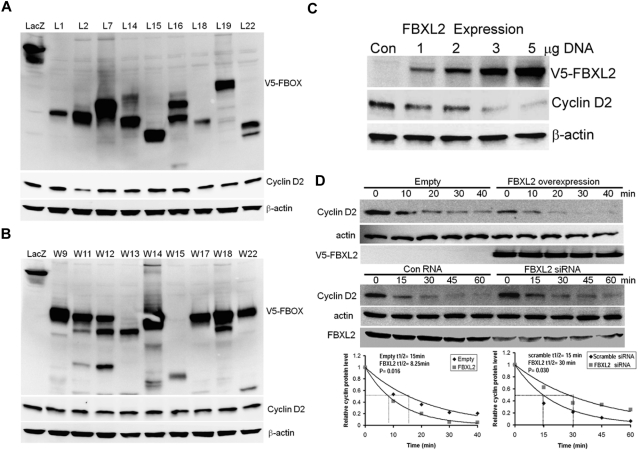
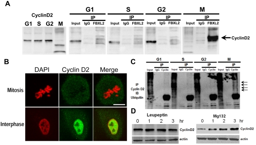
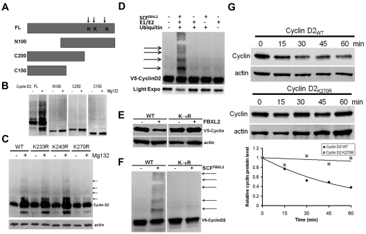
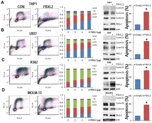
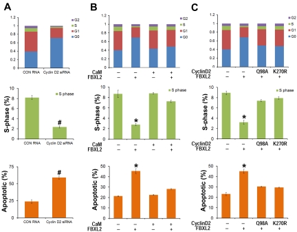
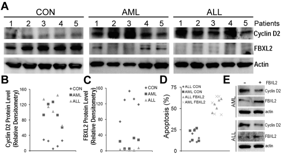
Hematologic maligancies exhibit a growth advantage by up-regulation of components within the molecular apparatus involved in cell-cycle progression. The SCF (Skip-Cullin1-F-box protein) E3 ligase family provides homeostatic feedback control of cell division by mediating ubiquitination and degradation of cell-cycle proteins. By screening several previously undescribed E3 ligase components, we describe the behavior of a relatively new SCF subunit, termed FBXL2, that ubiquitinates and destabilizes cyclin D2 protein leading to G(0) phase arrest and apoptosis in leukemic and B-lymphoblastoid cell lines. FBXL2 expression was strongly suppressed, and yet cyclin D2 protein levels were robustly expressed in acute myelogenous leukemia (AML) and acute lymphoblastic leukemia (ALL) patient samples. Depletion of endogenous FBXL2 stabilized cyclin D2 levels, whereas ectopically expressed FBXL2 decreased cyclin D2 lifespan. FBXL2 did not bind a phosphodegron within its substrate, which is typical of other F-box proteins, but uniquely targeted a calmodulin-binding signature within cyclin D2 to facilitate its polyubiquitination. Calmodulin competes with the F-box protein for access to this motif where it bound and protected cyclin D2 from FBXL2. Calmodulin reversed FBXL2-induced G(0) phase arrest and attenuated FBXL2-induced apoptosis of lymphoblastoid cells. These results suggest an antiproliferative effect of SCF(FBXL2) in lymphoproliferative malignancies.


Figures

Figure 1
Figure 1
Ectopic expression of FBXL2 induces the degradation of cyclin D2. (A-B) MLE cells were transfected with control plasmid lacZ or other V5 tagged F-box proteins (L and W families). Cells were collected and cell lysates were analyzed for V5, cyclin D2 and β-actin immunoblotting. (C) Cells were transfected with increasing amounts of V5-tagged FBXL2 plasmid. Cells were collected and cell lysates were analyzed for V5, cyclin D2, and β-actin immunoblotting. (D) Cyclin D2 protein half-life determination after FBXL2 overexpression (top panel), or FBXL2 knockdown using siRNA (middle panel). Bottom panel shows levels of cyclin D2 on immunoblots that were quantified densitometrically and are shown graphically. The data from each panel represent at least n = 2 separate experiments.
Figure 2
Figure 2
FBXL2 targets cyclin D2 for ubiquitination during mitosis. (A) MLE cells were synchronized to each cell phase followed by coimmunoprecipitation of endogenous FBXL2 and then cyclin D2 immunoblotting. (B) Cells were immunostained for cyclin D2 and counterstained with DAPI to visualize nucleus. (C) In vivo ubiquitination assays. Polyubiquitinated cyclin D2 was detected during cell-cycle progression by immunoprecipitation of endogenous cyclins followed by immunoblotting for ubiquitin. The arrows show polyubiquitinated cyclin D2. (D) Cyclin D2 levels in cells treated with leupeptin or MG132. The data from each panel represent at n = 2 separate experiments.
Figure 3
Figure 3
Cyclin D2 is polyubiquitinated at carboxyl-terminal acceptor sites. (A) Diagram of cyclin D2 deletion mutants constructed. Arrows indicate putative ubiquitin receptor sites. (B) V5-immunoblotting for levels of full-length (FL) or truncated cyclin D2 constructs after cellular expression and culture of cells in the absence (−) or presence (+) of MG132. (C) Cells were transfected with cyclin D2 point mutants before culture in the absence (−) or presence (+) of MG132. Immunoblotting was then performed for detection of accumulated (polyubiquitinated) cyclin D2 proteins in cells. The arrows indicate lack of polyubiquitinated signals after expression of the cyclin D2K270R construct. (D) In vitro ubiquitination assays. Purified SCF complex components were incubated with V5-cyclin D2 and the full complement of ubiquitination reaction components (second lane from left) showing polyubiquitinated cyclin D2. (E) Cyclin D2 protein levels in cells after cotransfection with either WT cyclin D2 or Lys270R cyclin D2 with or without ectopic FBXL2 expression. (F) In vitro ubiquitination assays. Purified SCF complex were incubated with WT V5-cyclin D2, or a Lys270R V5-cyclin D2 mutant and the full complement of ubiquitination reaction components. (G) Cyclin D2 protein half-life determination using cyclohexamide after expression of WT V5-cyclin D2, or Lys270R (V5-cyclin D2) mutant (data are from n = 2 experiments).
Figure 4
Figure 4
FBXL2 inhibits cell-cycle progression in transformed B-lymphoblastoid cells. (A) Immunoblotting showing levels of cyclins, CDKs, and negative control proteins, actin, and Erk after control (0) plasmid or ectopic FBXL2 expression. (B-C) FACS analysis in cells transfected with either empty plasmid or with increasing amount of FBXL2 plasmid. (D-E) Proliferation and viability studies of HCC1739 BL cells after FBXL2 overexpression. HCC1739 BL cells were transfected with FBXL2 for either 24 hours or 48 hours, cells were stained with trypan blue, and viable cells were analyzed by cell counting and graphed. n = 3 experiments, in control (C) #P < .01 versus empty plasmid, in panel D *P < .01 versus 0 hours, and in panel E *P < .01 versus control (CON).
Figure 5
Figure 5
FBXL2 induces G0 arrest in leukemic cell lines. (A-D) THP1, U937, K562, and MOLM-13 cells were cultured in RPMI medium followed by overexpression of FBXL2 using a GenomONE HVJ-E transfection kit. Cells were labeled with BrdU and analyzed by 2-color flow cytometry (left panels). Quantification of each cell phase is presented in the bar graphs (middle panels). Cells were also collected after transfection, cyclin Ds and FBXL2 protein levels were analyzed by immunoblotting (right immunoblots). Cells were also labeled with annexin V and analyzed by 2-color flow cytometry (far right panels). Apoptotic cells were quantified. *P < .05 versus empty plasmid.
Figure 6
Figure 6
FBXL2 and CaM differentially regulate the cell cycle in transformed B lymphoblastoid cells. (A) HCC1739 BL cells were transfected with either control RNA or cyclin D2 siRNA. Forty-eight hours later, cells were labeled with BrdU and analyzed through FACS or stained with annexin V and analyzed using a cell counter (n = 3 experiments, #P < 0.01 versus control (CON) RNA. (B) Cells were preinfected with a replication-deficient Adv-CaM or an empty virus and then transfected with FBXL2 plasmid. Forty-eight hours later, cells were either labeled with BrdU and analyzed through FACS or stained with annexin V and analyzed using a cell counter (n = 3 experiments, *P < .01 versus empty). (C) Cells were cotransfected with FBXL2 with or without 2 cyclin D2Q98A and cyclin D2K270R mutants. Forty-eight hours later, cells were either labeled with BrdU and analyzed through FACS or stained with annexin V and analyzed as previously described (n = 3 experiments, *P < .01 versus empty).
Figure 7
Figure 7
FBXL2 and cyclin D2 are differentially expressed in AML and ALL subjects. (A-C) PBMCs from 5 controls, AML, and ALL subjects were cultured in RMPI medium for 18 hours. Cells were then collected, lysed, and assayed for FBXL2 and cyclin D2 by immunoblotting (A). FBXL2 and cyclin D2 protein levels were quantified by densiometry using ImageJ Version 1.45 software, and the distribution of cyclin D2 (B) and FBLX2 protein (C) in each individual subject was graphed (n = 5, P < .005 vs control). (D) Leukemic cells were transfected with FBXL2 using a HVJ-E transfection kit for 24 hours. Cells were stained with annexin V and processed by flow cytometry. Apoptotic cells were quantified and graphed. (n = 5, P < .01 vs control). (E) FBXL2 and cyclin D2 protein levels were also analyzed from representative of AML and ALL transfected PBMCs.

References

    1. Buchakjian MR, Kornbluth S. The engine driving the ship: metabolic steering of cell proliferation and death. Nat Rev Mol Cell Biol. 2010;11(10):715–727. - PubMed
    1. Siebert R, Willers CP, Opalka B. Role of the cyclin-dependent kinase 4 and 6 inhibitor gene family p15, p16, p18, and p19 in leukemia and lymphoma. Leuk Lymphoma. 1996;23(5-6):505–520. - PubMed
    1. Ohtani K, DeGregori J, Nevins JR. Regulation of the cyclin E gene by transcription factor E2F1. Proc Natl Acad Sci U S A. 1995;92(26):12146–12150. - PMC - PubMed
    1. Lew DJ, Dulic V, Reed SI. Isolation of three novel human cyclins by rescue of G1 cyclin (Cln) function in yeast. Cell. 1991;66(6):1197–1206. - PubMed
    1. Gautschi O, Ratschiller D, Gugger M, Betticher DC, Heighway J. Cyclin D1 in non-small cell lung cancer: a key driver of malignant transformation. Lung Cancer. 2007;55(1):1–14. - PubMed

Publication types

MeSH terms