. 2012 Feb 21;109(8):3149-54.
doi: 10.1073/pnas.1120195109. Epub 2012 Feb 8.

Small molecule antagonist reveals seizure-induced mediation of neuronal injury by prostaglandin E2 receptor subtype EP2

Affiliations

Small molecule antagonist reveals seizure-induced mediation of neuronal injury by prostaglandin E2 receptor subtype EP2

Jianxiong Jiang et al. Proc Natl Acad Sci U S A. .

Abstract

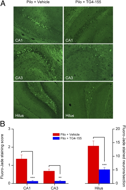
With interest waning in the use of cyclooxygenase-2 (COX-2) inhibitors for inflammatory disease, prostaglandin receptors provide alternative targets for the treatment of COX-2-mediated pathological conditions in both the periphery and the central nervous system. Activation of prostaglandin E2 receptor (PGE(2)) subtype EP2 promotes inflammation and is just beginning to be explored as a therapeutic target. To better understand physiological and pathological functions of the prostaglandin EP2 receptor, we developed a suite of small molecules with a 3-aryl-acrylamide scaffold as selective EP2 antagonists. The 12 most potent compounds displayed competitive antagonism of the human EP2 receptor with K(B) 2-20 nM in Schild regression analysis and 268- to 4,730-fold selectivity over the prostaglandin EP4 receptor. A brain-permeant compound completely suppressed the up-regulation of COX-2 mRNA in rat cultured microglia by EP2 activation and significantly reduced neuronal injury in hippocampus when administered in mice beginning 1 h after termination of pilocarpine-induced status epilepticus. The salutary actions of this novel group of antagonists raise the possibility that selective block of EP2 signaling via small molecules can be an innovative therapeutic strategy for inflammation-related brain injury.


Conflict of interest statement

The authors declare no conflict of interest.

Figures

Fig. 1.
Fig. 1.
Selective inhibition of EP2 receptor by hit compounds. (A) Chemical structures of TG4-155 and TG4-166. (B) TG4-155 and TG4-166 caused rightward shifts in the PGE2 dose–response curves in C6G-EP2 cells. TG4-155 (1 μM) caused a 1,120-fold shift and TG4-166 (1 μM) caused a 651-fold shift in the PGE2 EC50. (C) TG4-155 (1 μM) caused a 962-fold shift and TG4-166 (1 μM) caused a 678-fold shift in the butaprost EC50 in C6G-EP2 cells. (D) TG4-155 and TG4-166 (1 μM) had no effect on prostaglandin EP4 receptor. At 10 μM, TG4-155 caused only a 1.8-fold shift and TG4-166 caused an 8.6-fold shift in the PGE2 EC50 in HEK-EP4 cells. (E) There was no effect of TG4-155 and TG4-166 (10 μM) on β2-adrenergic receptor as shown by overlapping isoproterenol dose–response curves. Data were normalized as percentage of maximum response; points represent mean ± SEM (n = 4).
Fig. 2.
Fig. 2.
Competitive antagonism of EP2 receptor. (AC) Hits TG4-155, TG4-166, and analog TG4-292-1 inhibited PGE2-induced human EP2 receptor activation in a concentration-dependent manner. (D) Schild regression analysis was performed to elucidate the modality of antagonism from these compounds. TG4-155, TG4-166, and TG4-292-1 displayed a competitive antagonism mode of action on EP2 receptor shown by Schild plots. KB = 2.4, 4.6, and 1.8 nM; slopes, 1.0, 1.1, and 1.2 for TG4-155, TG4-166, and TG4-292-1, respectively. (E) EP2 antagonist compounds showed low potency on human EP4 receptor in HEK-EP4 cells, as illustrated by TG4-166. (F) Schild regression analysis was performed to evaluate inhibition of human EP4 receptor by TG4-166. KB = 2 μM; slope, 1.5. Data were normalized as percentage of maximum response; points represent mean ± SEM (n = 4).
Fig. 3.
Fig. 3.
Hit structure and analog design. Analogs were designed on the basis of the structure of hits TG4-155 and TG4-166.
Fig. 4.
Fig. 4.
EP2 activation induces microglial activation. (A) Butaprost increased cAMP levels in rat primary microglial cultures with EC50 = 0.5 μM. TG4-155 showed robust inhibition of butaprost-induced cAMP accumulation in rat microglia in a concentration-dependent manner, with potency equivalent to a Schild KB = 5 nM. (B) EP2 activation in microglia by butaprost (1 μM) did not affect expression of the EP2 receptor itself in cultured microglia. (C) Butaprost (1 μM) induced COX-2 expression 16.3-fold above background in microglia, measured by quantitative real-time PCR (qRT-PCR). The COX-2 up-regulation was attenuated by TG4-155. (D) BW245C, a selective DP1 agonist, did not induce microglial COX-2 at 10 nM. Bars represent the mean ± SEM (n = 3–4). ***P < 0.001 by one-way ANOVA with posthoc Bonferroni.
Fig. 5.
Fig. 5.
EP2 inhibition reduced neuronal injury after SE. (A) Neurodegeneration in hippocampi from animals treated with vehicle or TG4-155 was assessed by Fluoro-Jade staining 24 h after SE. No positive staining was detected in control mice treated with vehicle or TG4-155. (B) Quantification of neurodegenerating neurons in hippocampal subregions CA1, CA3, and dentate hilus. Coronal brain sections from 10 animals in each group were examined with a fluorescence microscope. Neuron injury in CA1 and CA3 was quantified by averaging the injury scores from three to nine sections per mouse (in each section: 0, <3 Fluoro-Jade–positive cells; 1, 3–30 cells; 2, 31–100 cells; 3, extensive Fluoro-Jade staining), exemplified in Fig. S9. Neuron injury in the hilus was evaluated by counts of Fluoro-Jade–positive cells per section (n = 10 mice per group, **P < 0.01, ***P < 0.001; one-way ANOVA and posthoc Bonferroni with selected pairs for CA1 and CA3, t test for hilus). Data are shown as mean ± SEM.

References

    1. Kaufmann WE, Worley PF, Pegg J, Bremer M, Isakson P. COX-2, a synaptically induced enzyme, is expressed by excitatory neurons at postsynaptic sites in rat cerebral cortex. Proc Natl Acad Sci USA. 1996;93:2317–2321. - PMC - PubMed
    1. Marcheselli VL, Bazan NG. Sustained induction of prostaglandin endoperoxide synthase-2 by seizures in hippocampus. Inhibition by a platelet-activating factor antagonist. J Biol Chem. 1996;271:24794–24799. - PubMed
    1. McCullough L, et al. Neuroprotective function of the PGE2 EP2 receptor in cerebral ischemia. J Neurosci. 2004;24:257–268. - PMC - PubMed
    1. Nakayama M, et al. Cyclooxygenase-2 inhibition prevents delayed death of CA1 hippocampal neurons following global ischemia. Proc Natl Acad Sci USA. 1998;95:10954–10959. - PMC - PubMed
    1. Iadecola C, et al. Reduced susceptibility to ischemic brain injury and N-methyl-D-aspartate-mediated neurotoxicity in cyclooxygenase-2-deficient mice. Proc Natl Acad Sci USA. 2001;98:1294–1299. - PMC - PubMed

Publication types

MeSH terms

Substances