. 2012 May;107(9):2342-51.
doi: 10.1152/jn.00973.2011. Epub 2012 Feb 8.

Attentional selection of location and modality in vision and touch modulates low-frequency activity in associated sensory cortices

Affiliations

Attentional selection of location and modality in vision and touch modulates low-frequency activity in associated sensory cortices

Markus Bauer et al. J Neurophysiol. 2012 May.

Abstract

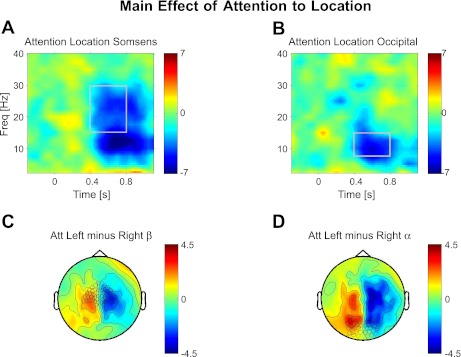
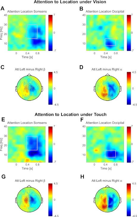
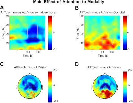
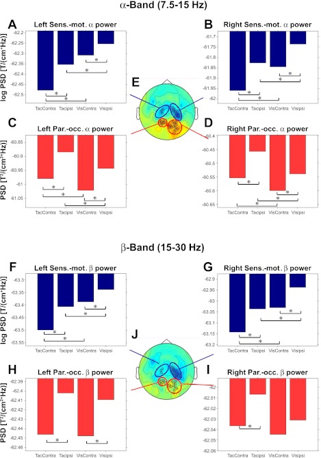
Selective attention allows us to focus on particular sensory modalities and locations. Relatively little is known about how attention to a sensory modality may relate to selection of other features, such as spatial location, in terms of brain oscillations, although it has been proposed that low-frequency modulation (α- and β-bands) may be key. Here, we investigated how attention to space (left or right) and attention to modality (vision or touch) affect ongoing low-frequency oscillatory brain activity over human sensory cortex. Magnetoencephalography was recorded while participants performed a visual or tactile task. In different blocks, touch or vision was task-relevant, whereas spatial attention was cued to the left or right on each trial. Attending to one or other modality suppressed α-oscillations over the corresponding sensory cortex. Spatial attention led to reduced α-oscillations over both sensorimotor and occipital cortex contralateral to the attended location in the cue-target interval, when either modality was task-relevant. Even modality-selective sensors also showed spatial-attention effects for both modalities. The visual and sensorimotor results were generally highly convergent, yet, although attention effects in occipital cortex were dominant in the α-band, in sensorimotor cortex, these were also clearly present in the β-band. These results extend previous findings that spatial attention can operate in a multimodal fashion and indicate that attention to space and modality both rely on similar mechanisms that modulate low-frequency oscillations.


Figures

Fig. 1.
Fig. 1.
Task schematics. A: experimental task setup. Participants fixate centrally, peripheral stimuli are presented to left or right, with tactile (Tac.) and visual (Vic.) stimuli in close spatial proximity. B: timeline: each trial starts with the presentation of a central cue for 100 ms, followed by a 700-ms empty interval, then presentation of either a tactile stimulus (0.5-ms electrical shock) or visual stimulus [light-emitting diode (LED) 200 ms]. Note that our magnetoencephalography (MEG) analyses focus on the cue-target interval, before target stimulus, to highlight pure top-down effects of preparatory attention (in absence of peripheral target stimulus-related effects). C: mapping of the visual or tactile (target) stimulus-induced responses (β-band suppression, 15–30 Hz) on the planar gradients. Left tactile stimuli suppress β-activity in predominantly right sensorimotor channels, and right tactile stimuli in left sensorimotor channels. Visual stimuli suppress β-activity in bilateral occipital channels. Note the spatial specificity of these response patterns. These sensors are used and labeled as sensorimotor and occipital channels throughout Figs. 2–4.
Fig. 2.
Fig. 2.
Attention to spatial location (main effect). Topographies and time-frequency (Freq) representations of the spatial-attention effect (regardless of modality judged). A and B: time-frequency plot of contralateral attention effects: (AttLeft−AttRight)left sensors minus (AttLeft−AttRight)right sensors. A: plots for the sensors over somatosensory cortex highlighted in C, whereas B shows this for the sensors over occipital cortex highlighted in D. Shown are the topography of t-values for attending left minus right in the β-band (C) of 15–30 Hz (A) and the α-band (D) of 7.5–15 Hz (B) over the time-frequency windows marked in A and B above, respectively. Topographies are averaged over the time-frequency windows as marked in A for C and B for D. Sensor selection is marked in C and D for time-frequency plots in A and B, respectively. All plots show t-values.
Fig. 3.
Fig. 3.
Attention to spatial location under vision and touch. Topographies and time-frequency representations of spatial-attention effects are shown analogously to Fig. 2 but now separately for vision or touch being task-relevant. A–D: spatial-attention effect when vision-relevant. A and B: time-frequency plot of contralateral attention effects: (AttLeft−AttRight)left sensors minus (AttLeft−AttRight)right sensors for sensorimotor (A) and occipital (B) sensors marked in C and D, respectively. C and D: topography of t-values attending left minus right for β- (C) and α- (D) activity, averaged over time-frequency windows marked in A and B, respectively. E–H: attention to space when touch-relevant. E and F: time-frequency plot of contralateral attention effects: (AttLeft−AttRight)left sensors minus (AttLeft−AttRight)right sensors for sensorimotor (E) and occipital (F) sensors marked in G and H, respectively. G and H: topography of t-values attending left minus right for β- (G) and α- (H) activity, averaged over time-frequency windows marked in E and F, respectively. All plots show t-values.
Fig. 4.
Fig. 4.
Attention to modality. Topographies and time-frequency representations of modality relevance effect (regardless of attended side). A and B: time-frequency plot of α- and β-suppression for somatosensory (A) and occipital (B) sensors; sensor selection is marked in C and D, respectively. All plots show t-values. C and D: topography of attending touch minus vision for β- (C) and α- (D) bands (B), averaged over time-frequency windows marked in A and B, respectively.
Fig. 5.
Fig. 5.
Attention to space and modality at modality-selective sensors. Activity profiles for α-activity (7.5–15 Hz, for 0.4–0.8 s into the cue-target interval; A–D) at sensors selected to show a preference (significance exceeding P < 0.01) either for touch being task-relevant, which arose at sensorimotor sensors (Sens.-mot.; A and B), or for vision being task-relevant, which arose at parieto-occipital sensors (Par.-occ.; C and D) as marked in E, the middle head view (occipital red and sensorimotor blue), which also depicts the topography for the contrast “Attend Touch” minus “Attend Vision” (in the α-band). The bar plots show the mean log-transformed α-power of magnetic induction in planar gradients (hence negative log values) for the 4 different attentional conditions (“Touch Left,” “Touch Right,” “Vision Left,” and “Vision Right”) in the preparatory cue-target interval. The conditions are plotted relative to the lateral sensor positions (Touch Right becomes “TacContra” for left sensors). Asterisks indicate significant (P < 0.05) attention to modality and location effects (by paired t-test, which are found for every case). In particular, note that spatial-attention effects are significant even in sensors that show a clear preference for the other modality. H–J: same as above (A–E) but now for the β-band (15–30 Hz, for 0.4–0.8 s into the cue-target interval). Whereas all contrast in sensorimotor sensors are also significant for the β-band, in parieto-occipital sensors, they were not. Since effects of modality selection in the β-band were not significant anywhere in sensors over parieto-occipital cortex, the sensor selection (J) here was therefore adopted from the contrast of attention to touch minus vision in the α-band (E). PSD, power spectral density; T, time; ipsi, ipsilateral; Contra, contralateral.

References

    1. Banerjee S, Snyder AC, Molholm S, Foxe JJ. Oscillatory alpha-band mechanisms and the deployment of spatial attention to anticipated auditory and visual target locations: supramodel or sensory-specific control mechanisms? J Neurosci 31: 9923–9932, 2011 - PMC - PubMed
    1. Bastiaansen MC, Knösche TR. Tangential derivative mapping of axial MEG applied to event-related desynchronization research. Clin Neurophysiol 111: 1300–1305, 2000 - PubMed
    1. Bauer M, Oostenveld R, Peeters M, Fries P. Tactile spatial attention enhances gamma-band activity in somatosensory cortex and reduces low-frequency activity in parieto-occipital areas. J Neurosci 26: 490–501, 2006 - PMC - PubMed
    1. Buffalo EA, Fries P, Landman R, Liang H, Desimone R. A backward progression of attentional effects in the ventral stream. Proc Natl Acad Sci USA 107: 361–365, 2010 - PMC - PubMed
    1. Donner TH, Siegel M, Oostenveld R, Fries P, Bauer M, Engel AK. Population activity in the human dorsal pathway predicts the accuracy of visual motion detection. J Neurophysiol 98: 345–359, 2007 - PubMed

Publication types

LinkOut - more resources