Comparative Study
. 2012 Feb 8;32(6):1974-80.
doi: 10.1523/JNEUROSCI.5054-11.2012.

Coding of microsaccades in three-dimensional space by premotor saccadic neurons

Affiliations
Comparative Study

Coding of microsaccades in three-dimensional space by premotor saccadic neurons

Marion R Van Horn et al. J Neurosci. .

Abstract

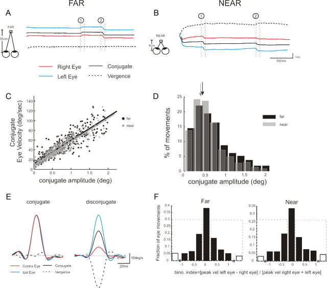
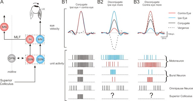
Microsaccades are small, involuntary eye movements that are produced during fixation. While accurate visual perception requires precise binocular coordination during fixation, previous studies of the neural control of microsaccades measured the movement of one eye only. Here we show how premotor saccadic neurons control these small fixational eye movements in three-dimensional space. Microsaccadic eye movements, produced by monkeys trained to fixate targets presented at different depths, were similarly distributed in three-dimensional space during both near and far viewing. Single unit recordings of the neural activity of premotor neurons further revealed that the brainstem saccadic circuitry controls these minute disconjugate shifts of gaze by preferentially encoding the dynamic movement of an individual eye (i.e., integrated control of conjugate and vergence motion). These findings challenge the traditional notion that microsaccades are strictly conjugate and have important implications for studies that use microsaccades to evaluate visual and attentional processing, as well as certain neurological disorders.


Figures

Figure 1.
Figure 1.
A, B, Example traces of the eye position during fixation of a far (A) and near (B) target. C, Peak velocity–amplitude relationship for microsaccades during near (gray dots; N = 365) and far (black dots; N = 362) fixation. D, Distribution of the size of microsaccades detected during near (gray) and far (black) fixation. E, Example velocity profiles of a conjugate (left) and disconjugate (right) microsaccade. F, Distribution of the degree of conjugacy across all microsaccades during far and near viewing. Open bars denote movements where the eyes moved in opposite directions. Dashed gray boxes denote microsaccades where the movement of eye each differed by >20%. Contra, Contralateral; Ipsi, ipsilateral; bino., binocular; vel, velocity.
Figure 2.
Figure 2.
A, Example neural activity from a typical SBN (left) and OPN (right) during conjugate microsaccades aligned on saccade onset. Average firing rate with standard deviations is superimposed on the unit activity. Average ipsilateral (Ipsi), contralateral (Contra), conjugate, and vergence velocities traces are shown below the raster plots. B, Example raster plots on the same example SBN shown in A during disconjugate saccades where the ipsilateral (left) or contralateral (right) eye moved more. C, Bootstrap histograms and 95% confidence intervals (thick horizontal bars) for example SBN.
Figure 3.
Figure 3.
A, B, Distribution of ratio indexes for SBNs (A) and motoneurons (B) (see Materials and Methods). Insets show distributions of ratios for a sample of SBNs (left) and motoneurons (right) estimated in previous studies (Van Horn and Cullen, 2008). MONO, Monocular; BINO, binocular; CONTRA, contralateral; IPSI, ipsilateral; CONJ, conjugate.
Figure 4.
Figure 4.
A, Illustration of the neural circuitry involved in generating microsaccades. The superior colliculus projects to the SBNs, which are reciprocally connected with the OPNs, and project directly to the motoneurons (MN) and internuclear neurons (IN) in the abducens, which project to the contralateral abducens via the medial longitudinal fasicularis (MLF). Neurons encoding the movement of the ipsilateral (Ipsi) and contralateral (Contra) eye are shown in blue and red, respectively. B, Example unit activity of premotor neurons during example conjugate (B1) and disconjugate microsaccades (B2, B3). Blue unit activity is the activity associated with a neuron related to the movement of the ipsilateral eye and red unit activity is representative of a neuron related to the movement of the contralateral eye. LR, Lateral rectus.

References

    1. Betta E, Turatto M. Are you ready? I can tell by looking at your microsaccades. Neuroreport. 2006;17:1001–1004. - PubMed
    1. Brien DC, Corneil BD, Fecteau JH, Bell AH, Munoz DP. The behavioural and neurophysiological modulation of microsaccades in monkeys. J Eye Mov Res. 2009;3:1–12.
    1. Carpenter J, Bithell J. Bootstrap confidence intervals: when, which, what? A practical guide for medical statisticians. Stat Med. 2000;19:1141–1164. - PubMed
    1. Cornsweet TN. Determination of the stimuli for involuntary drifts and saccadic eye movements. J Opt Soc Am. 1956;46:987–993. - PubMed
    1. Cui J, Wilke M, Logothetis NK, Leopold DA, Liang H. Visibility states modulate microsaccade rate and direction. Vision Res. 2009;49:228–236. - PMC - PubMed

Publication types

LinkOut - more resources