. 2012 Mar 23;335(6075):1492-6.
doi: 10.1126/science.1218091. Epub 2012 Feb 9.

Plant UVR8 photoreceptor senses UV-B by tryptophan-mediated disruption of cross-dimer salt bridges

Affiliations

Plant UVR8 photoreceptor senses UV-B by tryptophan-mediated disruption of cross-dimer salt bridges

John M Christie et al. Science. .

Abstract

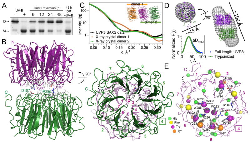
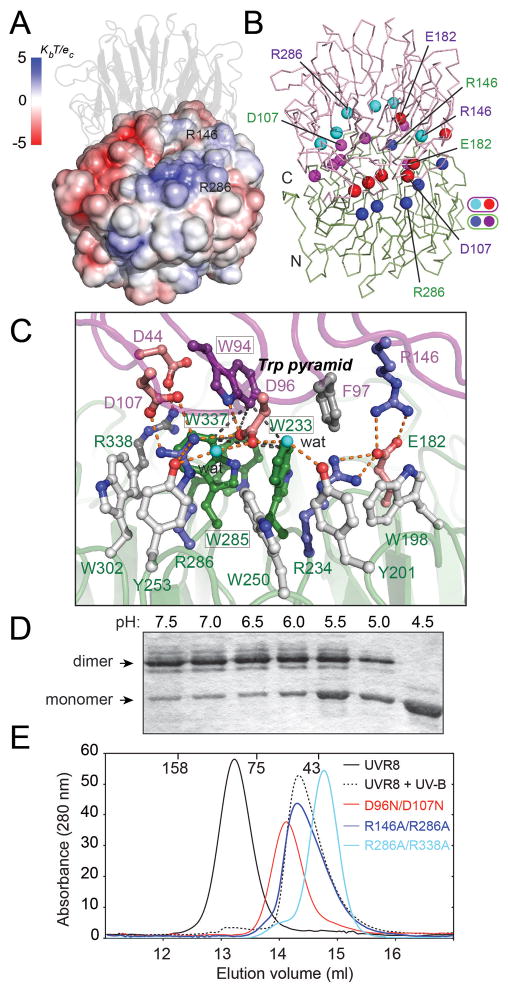
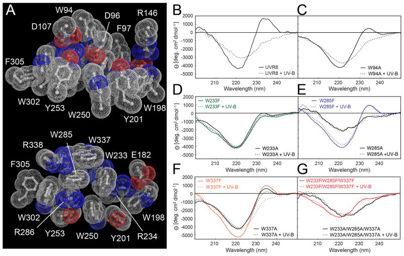
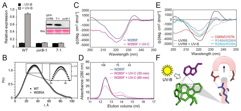
The recently identified plant photoreceptor UVR8 (UV RESISTANCE LOCUS 8) triggers regulatory changes in gene expression in response to ultraviolet-B (UV-B) light through an unknown mechanism. Here, crystallographic and solution structures of the UVR8 homodimer, together with mutagenesis and far-UV circular dichroism spectroscopy, reveal its mechanisms for UV-B perception and signal transduction. β-propeller subunits form a remarkable, tryptophan-dominated, dimer interface stitched together by a complex salt-bridge network. Salt-bridging arginines flank the excitonically coupled cross-dimer tryptophan "pyramid" responsible for UV-B sensing. Photoreception reversibly disrupts salt bridges, triggering dimer dissociation and signal initiation. Mutation of a single tryptophan to phenylalanine retunes the photoreceptor to detect UV-C wavelengths. Our analyses establish how UVR8 functions as a photoreceptor without a prosthetic chromophore to promote plant development and survival in sunlight.


Figures

Figure 1
Figure 1
Structure of Arabidopsis UVR8 dimer. (A) UV-B-induced dimer dissociation spontaneously reverses in the dark, regenerating photoactive dimers; analyzed by SDS-PAGE without sample boiling. (B) UVR8 forms a symmetric homo-dimer of 7-bladed β-propeller subunits (side-and end-views). Key salt bridges are shown as ball-and-stick. End-view is numbered to show blade pairing centered at blade 4 (box). (C) Experimental SAXS profile of UVR8 (crystallographic construct) compared with computed profiles for crystallographic dimers. (D) Crystallographic dimer docked into ab initio SAXS model of full-length UVR8 dimer (top) and pair-distance-distribution functions [P(r)] for full-length and trypsin-treated UVR8 (bottom), defining maximal diameter difference (ΔDmax). (E) Asymmetric localization of aromatic residues identifies center of photoreception. Key side chains (grey) are labeled.
Figure 2
Figure 2
Ionic interactions are key to maintaining the UVR8 dimer. (A) Electrostatic potential surface of UVR8 reveals charge complementarity at the dimer interface, (B) Arg (blue/cyan) and Glu/Asp (red/magenta) balls indicate key charged residues mediating cross-dimer salt bridges aligned across the interface. (C) Close up of key salt bridges: Arg 286 with Asp 96 and Asp 107; Arg 146 with Glu 182; Arg 338 with Asp 44, with hydrogen bonds shown as orange dots. The Trp pyramid (black dashed lines) is formed by W94 (purple) atop the Trp triad (green). (D) Acidification promotes monomerization of wild-type UVR8, as analyzed by SDS-PAGE without sample boiling (E) Size exclusion chromatography shows UVR8R146A/R286A, UVR8R286A/R338A and UVR8D96N/D107N mutants are constitutive monomers.
Figure 3
Figure 3
Specific tryptophans mediate UV-B photoreception by UVR8. (A) Bulky aromatic residues tightly pack with key charged residues across the dimer (top) and within a monomer (bottom). (B–G) Far-UV CD spectrum change of UVR8 by UV-B. (B) Wild-type UVR8; (C) UVR8W94A; (D) UVR8W233F and UVR8W233A; (E) UVR8W285F and UVR8W285A; (F) UVR8W337F and UVR8W337A; (G) triple mutants UVR8W233F/W285F/W337F and UVR8W233A/W285A/W337A.
Figure 4
Figure 4
Key features of UVR8 photoreceptor mechanism (A) qRT-PCR analysis of UV-B induction of HY5 transcripts in wild-type, uvr8-1, and uvr8-1 expressing GFP-UVR8W285A (line 7-1). Inset shows Western blot of GFP-UVR8 in plant lines and Ponceau S staining of rbcL protein as loading control. (B) SAXS Pair-distance-distribution functions [P(r)] show that UVR8W285A is dimeric, with only subtle conformational differences from wild-type. (C) Far-UV CD spectra of UVR8W285F before and after exposure to 40 min UV-C. (D) Size exclusion chromatography of UVR8W285F mutant protein exposed to UV-C (solid and dotted magenta lines). (E) Far-UV CD spectra of UVR8R146A/R286A, UVR8R286A/R338A and UVR8D96N/D107N mutants. (F) Model for UV-B photoreception by UVR8. UV-B sensing (by Trp “pyramid”) triggers dimer dissociation by disrupting salt bridges.

Comment in

References

    1. Brown BA, et al. A UV-B-specific signaling component orchestrates plant UV protection. Proc Natl Acad Sci U S A. 2005;102:18225. - PMC - PubMed
    1. Brown BA, Jenkins GI. UV-B signaling pathways with different fluence-rate response profiles are distinguished in mature Arabidopsis leaf tissue by requirement for UVR8, HY5, and HYH. Plant Physiol. 2008;146:576. - PMC - PubMed
    1. Favory JJ, et al. Interaction of COP1 and UVR8 regulates UV-B-induced photomorphogenesis and stress acclimation in Arabidopsis. EMBO J. 2009;28:591. - PMC - PubMed
    1. Jenkins GI. Signal transduction in responses to UV-B radiation. Annu Rev Plant Biol. 2009;60:407. - PubMed
    1. Kliebenstein DJ, Lim JE, Landry LG, Last RL. Arabidopsis UVR8 regulates ultraviolet-B signal transduction and tolerance and contains sequence similarity to human regulator of chromatin condensation 1. Plant Physiol. 2002;130:234. - PMC - PubMed

Publication types

MeSH terms

Associated data