. 2012 Mar 9;335(6073):1232-5.
doi: 10.1126/science.1217869. Epub 2012 Feb 9.

Triggering a cell shape change by exploiting preexisting actomyosin contractions

Affiliations

Triggering a cell shape change by exploiting preexisting actomyosin contractions

Minna Roh-Johnson et al. Science. .

Abstract

Apical constriction changes cell shapes, driving critical morphogenetic events, including gastrulation in diverse organisms and neural tube closure in vertebrates. Apical constriction is thought to be triggered by contraction of apical actomyosin networks. We found that apical actomyosin contractions began before cell shape changes in both Caenorhabitis elegans and Drosophila. In C. elegans, actomyosin networks were initially dynamic, contracting and generating cortical tension without substantial shrinking of apical surfaces. Apical cell-cell contact zones and actomyosin only later moved increasingly in concert, with no detectable change in actomyosin dynamics or cortical tension. Thus, apical constriction appears to be triggered not by a change in cortical tension, but by dynamic linking of apical cell-cell contact zones to an already contractile apical cortex.


Conflict of interest statement

The authors declare no conflicts of interest.

Figures
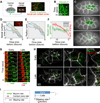
Figure 1
Figure 1. Actomyosin contraction precedes the rapid shrinking of the apical surface
(A) Diagram of imaging method. (B) NMY-2::GFP coalescence (white arrowheads) in apical cortex of Ea/p cell. (C) Shrinking of apical surfaces during gastrulation (projections of ten 1µm z-planes, with Ea/p false-colored). (D) Ea/p cell apical surface areas over 575 or 825 secs (5 embryos each) before closure of the apical surface. Inset: apical cell-cell contact zones (arrowheads) on Ep (asterisk). (E) Average radius of apical surfaces derived from area measurements. (F) Mean and 95%CI of radius and myosin particle rate over time. Inset: myosin directionality (net distance over total distance, vertical scale 0 to 1) over time (time scale: same as larger graph). (G) Movements of individual myosin particles (arrowheads) near contact zones (white dotted lines) in early or late stages of closure. Arrows at bottom: relative distances traveled by each. (H) PIV, three magnifications. Boxes indicate enlarged areas. Left to right: whole embryo at plane of Ea/p apical cortex, Ea/p cells (outlined by dotted line), part of Ea at border with another cell. (I) Slipping rate calculated from individual particles and contact zones (p<0.001, Student's t-test).
Figure 2
Figure 2. Periodic actomyosin coalescence occurs before apical cell profiles shrink in Drosophila gastrulation
(A) Drosophila ventral furrow formation. Circles mark apical myosin enrichment seen before apical cell profiles began to shrink. (B) Kymograph of a cell (diagrammed) showing myosin (green) movement toward a stationary cell-cell boundary (red) before apical shrinking began. (C) Myosin coalesced (green arrowheads) and dissipated (gray arrowheads) before apical cell profiles began to shrink. This is shown quantitatively from one cell in (D), and from 11 cells chosen at random in (E). Heatmaps in (E) show local maxima of apical myosin levels (3-timepoint running averages of myosin level at each timepoint minus the average of 10 timepoints before and after, normalized to maximum and minimum). Green and gray arrowheads mark one case as in (C). Cell 3 is a rare example where peaks were not seen before apical shrinking began. (F) Slipping rate, defined as in Fig. 1I, early (before apical shrinking, n=33 cells, 3 embryos) and late (during apical shrinking, n=27 cells, 3 embryos), p<0.01 (Student's t-test).
Figure 3
Figure 3. Cortical tension associated with apical constricton is established early and changes little as apical shrinking accelerates in C. elegans
(A) A spontaneous failure, with three timepoints overlain in three colors. Inset shows entire Ea/p cell apical cortex outlined with enlarged region indicated. Arrows mark individual myosin particles springing apart. (B) Similar data from Ea/p cortical laser cuts done in early or late stages by PIV as in Fig 1H. (C) Initial recoil speeds of myosin particles after spontaneous failures at early (n=13 myosin particles within 1µm of center of recoil, 6 embryos) and late (20 particles, 7 embryos) stages, or after laser cuts (48 particles within 4µm of cut site, 7 embryos/stage). Exponential decay T1/2 was 2.20 secs in early stages, n=12; 2.38 secs in late stages, n=20. (D) Working model of forces acting on contact zones (red) and within Ea/p apical actomyosin networks (green, with multiple, interconnected network elements represented as two elements here for simplicity). Results suggest that cortical tension (T) and network stiffness or viscous drag (green dashpots) within Ea/p change little from early to late stages.
Figure 4
Figure 4. Targeting classical cadherin and Rac signaling prevents coupled movements but not actomyosin contraction
(A) Closure speed (µm/min decrease in average diameter) of apical cell areas in hmr-1(RNAi) or ced-5(n1812) does not reach the speed found in wild-type embryos (asterisks: p<0.05). (B) PIV in hmr-1(RNAi);ced-5(n1812) doubles. Myosin moves centripetally with little membrane movement in the same direction at either stage. This is shown for individual particles in (D), with quantification as in Fig. 1I in (E). Black dotted lines on hmr-1 bars in (E) mark wild type for comparison. Asterisks: p<0.001 (Student's t-test).

Comment in

References

    1. Friedl P, Gilmour D. Collective cell migration in morphogenesis, regeneration and cancer. Nat Rev Mol Cell Biol. 2009;10(7):445–457. - PubMed
    1. Weijer CJ. Collective cell migration in development. J Cell Sci. 2009;122(Pt 18):3215–3223. - PubMed
    1. Odell GM, et al. The mechanical basis of morphogenesis. I. Epithelial folding and invagination. Dev Biol. 1981;85(2):446–462. - PubMed
    1. Sawyer JM, et al. Apical constriction: a cell shape change that can drive morphogenesis. Dev Biol. 2010;341(1):5–19. - PMC - PubMed
    1. Copp AJ, Greene ND. Genetics and development of neural tube defects. J Pathol. 2010;220(2):217–230. - PMC - PubMed

Publication types

MeSH terms