. 2012 Feb 10;335(6069):709-12.
doi: 10.1126/science.1214453.

Structure-based mechanistic insights into DNMT1-mediated maintenance DNA methylation

Affiliations

Structure-based mechanistic insights into DNMT1-mediated maintenance DNA methylation

Jikui Song et al. Science. .

Abstract

DNMT1, the major maintenance DNA methyltransferase in animals, helps to regulate gene expression, genome imprinting, and X-chromosome inactivation. We report on the crystal structure of a productive covalent mouse DNMT1(731-1602)-DNA complex containing a central hemimethylated CpG site. The methyl group of methylcytosine is positioned within a shallow hydrophobic concave surface, whereas the cytosine on the target strand is looped out and covalently anchored within the catalytic pocket. The DNA is distorted at the hemimethylated CpG step, with side chains from catalytic and recognition loops inserting through both grooves to fill an intercalation-type cavity associated with a dual base flip-out on partner strands. Structural and biochemical data establish how a combination of active and autoinhibitory mechanisms ensures the high fidelity of DNMT1-mediated maintenance DNA methylation.


Figures

Fig. 1
Fig. 1
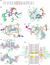
Structural overview of mDNMT1(731-1602) bound covalently to a hemi-mCpG site. (A) Ribbon representation of the covalent complex in two orthogonal views. The BAH1, BAH2, and methyltransferase domains are colored light pink, orange, and cyan, respectively; DNA and zinc ions are barley and purple, respectively. The disordered (Gly-Lys)n linker [(GK)n] is shown as black dashed lines, the 5-methyl group from mC6 is green, the flipped-out target fC7′ is purple, the flipped-out C from parental strand is blue, the catalytic loop and TRD loops 1 and 2 are green, and the bound AdoHcy is in a space-filling representation. (B) Structural superposition of the covalent mDNMT1(731-1602)–DNA complex with the autoinhibited mDNMT1(650-1602)–DNA complex. The bound DNA in the covalent complex is in beige with the protein in cyan; they are yellow and cyan in the autoinhibited complex. The catalytic loop (expanded view in inset) is colored purple in the autoinhibited complex and green in the covalent complex. The CXXC-BAH1 domain linker is in dark blue in the autoinhibited complex. The inset highlights the transition from a straight helix (green) in the productive covalent complex to a kinked helix (purple) in the autoinhibited complex. (C) Close-up view of the catalytic loop (in green) in the covalent mDNMT1 (731-1602)–DNA complex (this study). (D) Close-up view of the catalytic loop (in dark brown) in the autoinhibited mDNMT1(650-1602)–DNA complex (PDB: 3PT6). The side chain of residue Cys1229 is shown in stick representation.
Fig. 2
Fig. 2
Insertion of amino acid side chains into the intercalation-like distortion of the bound DNA and base-specific recognition at the hemi-mCpG site in the covalent mDNMT1-DNA complex. (A) Sequencing and numbering system of the hemi-mCpG–containing 12-mer duplex. (B) Stabilization of the flipped-out target cytosine fC7′ (light blue ball) in the active site of the enzyme. (C) The 5-methyl group (green ball) of mC6 is anchored within a hydrophobic concave surface of mDNMT1. (D) Insertion of side chains of Met1235 and Lys1537 into the intercalation-type space, and buttressing by indole ring of Trp1512. The covalent bond between the sulfur atom of Cys1229 and the C6 atom of the target cytosine fC7′ is shown as a dark line. (E) End-on view of Met1235 and Lys1537 side chain insertion in the covalent complex. (F) Direct and water-mediated (labeled with purple W) hydrogen bond interactions target the Watson-Crick mC6-G6′ base pair from both grooves. (G) Stabilization of the repositioned guanine G7 through hydrogen-bonding interactions. (H) Schematic tabulation of direct and water-mediated hydrogen bond and electrostatic interactions between mDNMT1 and DNA in the covalent complex. Abbreviations for amino acids: C, Cys; E, Glu; F, Phe; G, Gly; H, His; K, Lys; L, Leu; M, Met; N, Asn; P, Pro; Q, Gln; R, Arg; S, Ser; T, Thr; W, Trp; Y, Tyr.
Fig. 3
Fig. 3
Insertion of the catalytic loop and a pair of TRD loops into the grooves centered about the hemi-mCpG site in the covalent mDNMT1-DNA complex. (A) Catalytic loop–DNA interactions. (B) TRD loop 1–DNA interactions. The imidazole ring of His1504 bridges the zinc finger with the backbone phosphate of mC6. (C) TRD loop 2–DNA interactions. (D) Intermolecular hydrogen-bond interactions between side chains of a segment of the BAH2 loop and DNA in the complex. (E) Methylation activities of mDNMT1(731-1602) and its mutants involving amino acids that line the mC-recognizing hydrophobic surface, or insert into the intercalation-type cavity, and catalytic and BAH2 loop residues involved in substrate recognition. Methylation activities were monitored after reaction on hemi-mCpG DNA substrate (black bars) or unmodified CpG DNA substrate (white bars) for 10 min. Error bars represent SD calculated from two measurements.

References

    1. Cheng X, Blumenthal RM. Structure. 2008;16:341. - PMC - PubMed
    1. Goll MG, Bestor TH. Annu. Rev. Biochem. 2005;74:481. - PubMed
    1. Law JA, Jacobsen SE. Nat. Rev. Genet. 2010;11:204. - PMC - PubMed
    1. Suzuki MM, Bird A. Nat. Rev. Genet. 2008;9:465. - PubMed
    1. Li E, Bestor TH, Jaenisch R. Cell. 1992;69:915. - PubMed

Publication types

MeSH terms

Associated data