Review
. 2011 Dec;1(6):476-86.
doi: 10.1016/j.coviro.2011.11.001.

Induction and function of type I and III interferon in response to viral infection

Affiliations
Review

Induction and function of type I and III interferon in response to viral infection

David E Levy et al. Curr Opin Virol. 2011 Dec.

Abstract

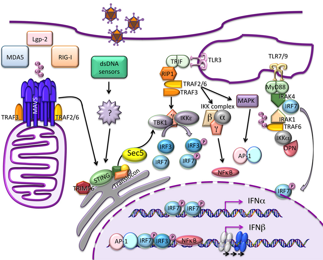
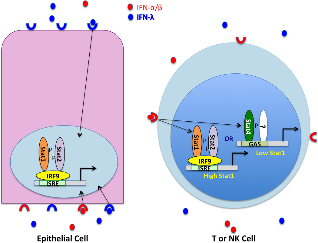
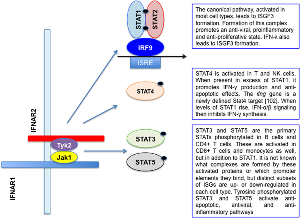
The type I and III interferon (IFN) families consist of cytokines rapidly induced during viral infection that confer antiviral protection on target cells and are critical components of innate immune responses and the transition to effective adaptive immunity. The regulation of their expression involves an intricate and stringently regulated signaling cascade, initiated by recognition most often of viral nucleic acid in cytoplasmic and endosomal compartments and involving a series of protein conformational rearrangements and interactions regulated by helicase action, ubiquitin modification, and protein aggregation, culminating in kinase activation and phosphorylation of critical transcription factors and their regulators. The many IFN subtypes induced by viruses confer amplification, diversification, and cell-type specificity to the host response to infection, providing fertile ground for development of antiviral therapeutics and vaccines.


Figures

Figure 1
Figure 1. Induction IFN by viruses
Virus infection triggers a complex signaling cascade involving ubiquitin modification and protein aggregation, leading to kinase activation and transcription factor phosphorylation. Signaling is initiated by the detection of viral nucleic acid by both cytoplasmic helicase receptors and endosomal transmembrane Toll-like receptors, activating distinct signaling pathways that converge on transcription factor phosphorylation. Kinase activation is controlled by signaling protein aggregates that assemble as scaffolds on organellar surfaces, including mitochondria, endoplasmic reticulum, and possibly peroxisomes. Phosphorylated IRF3 and IRF7 and activated NF-κB translocate to the cell nucleus, where they bind to enhancer/promoter regions of IFN genes. IFN-β (and probably some IFN-λ subtypes) requires assembly of a multiprotein enhanceosome that alters chromatin structure, sliding an occluding nucleosome that would otherwise prevent polymerase recruitment. The multiple type I and III IFN genes are largely regulated in analogous fashions, although differential utilization of multiple IRF isoforms, some of which are subject to feedforward and feedback regulation, produces distinct patterns of induction of the individual genes under different circumstances.
Figure 2
Figure 2. Tissue-specific biological effects of type I and III IFN
IFN-α, -β and -λ are induced by virus infection and secreted by both infected cells and plasmacytoid DCs. Their relative ratio may vary with the pathogen, but IFN-λ is the predominant cytokine secreted by either cell type in the course of influenza virus infection. IFNs can be detected in the blood and in bronchoalveolar lavage fluid during respiratory infection. Respiratory and intestinal epithelial cells are polarized, and there is evidence that IFN receptor expression on intestinal epithelia is polarized as well, with IFNAR localized to basolateral surfaces and IFNLR present on both basolateral and apical surfaces. Receptors on the basolateral surface can detect IFNs present in blood, while apical receptors will see IFNs present at the luminal surface. Signaling through IFNAR or the IFN-λ receptor leads to Stat1 and Stat2 tyrosine phosphorylation, and formation of the ISGF3 complex consisting of phospho-Stat1, phospho-Stat2 and IRF9, which activates target gene expression following nuclear translocation. T and NK cells lack the IFNLR chain, and therefore cannot respond to IFN-λ. However, the response to signaling through IFNAR is conditional (see also Fig. 3). In naïve mice, both Stat1 and Stat4 are present in NK and T cells, with constitutive levels of Stat1 much lower than Stat4. In this state, IFN-α/β signaling results predominantly in Stat4 phosphorylation, which occurs in a Stat2 independent manner and triggers IFN-γ production. In the course of viral infection, Stat1 levels are induced by IFN signaling, altering the ratio of Stat1 to Stat4 in stimulated cells. The consequences of this shift are only partly understood, but serve to explain seemingly contradictory responses of T and NK cells to IFN-α treatment. For example, despite the known antiproliferative effect of IFN-α, virus specific CD8 T cell expansion can occur at a time when IFN levels are high. However, low levels of Stat1 in actively dividing CD8 T cells from infected mice render this population resistant to the anti-proliferative effects of IFN.
Figure 3
Figure 3. Cell type-specific biological effects of type I IFN during virus infection
IFN-α, and -β are induced by virus infection and secreted by both infected cells and plasmacytoid DCs. In most cell types, signaling through IFNAR receptor leads to Stat1 and Stat2 tyrosine phosphorylation, and formation of the canonical ISGF3 complex consisting of phospho-Stat1, phospho-Stat2 and IRF9, which activates target gene expression following nuclear translocation. However, the response to signaling through IFNAR is both cell specific and context dependent. In most cells studied Stat3 is also activated by IFN-αβ, and acts as a brake on Stat1-mediated, antiviral, proinflammatory, proapoptotic gene expression. Both Stat1 and Stat4 are activated in NK and CD4+ T cells, but Stat4-mediated IFN-γ production by these cells requires low levels of Stat1. Stats1, 3 and 5 are activated by IFN-β treatment of resting B and T cells, with a cell type specific pattern, with very little Stat1 activation in B cells and CD4+ T cells. Stat5, like Stat3, is anti-apoptotic and promotes cell proliferation. However, even in the absence of pStat1, antiviral ISGs can be expressed through IRF-containing complexes binding to alternative enhancer elements [88]. The extent to which IFN-λ activates Stats other than Stat1 and Stat2 has yet to be determined.

References

    1. Donnelly RP, Kotenko SV. Interferon-lambda. a new addition to an old family. J Interferon Cytokine Res. 2010;30:555–564. - PMC - PubMed
    1. Iversen MB, Paludan SR. Mechanisms of type III interferon expression. J Interferon Cytokine Res. 2010;30:573–578. - PubMed
    1. Stetson DB, Medzhitov R. Type I interferons in host defense. Immunity. 2006;25:373–381. - PubMed
    1. Lin WJ, Zheng X, Lin CC, Tsao J, Zhu X, Cody JJ, Coleman JM, Gherzi R, Luo M, Townes TM, et al. Posttranscriptional control of type I interferon genes by KSRP in the innate immune response against viral infection. Mol Cell Biol. 2011;31:3196–3207. Documents a new mechanism for translational control of type I IFN synthesis.

    1. Marie I, Durbin JE, Levy DE. Differential viral induction of distinct interferon-alpha genes by positive feedback through interferon regulatory factor-7. EMBO J. 1998;17:6660–6669. - PMC - PubMed