The single incision laparoscopic intragastric wedge resection of gastric submucosal tumor
- PMID: 22324014
- PMCID: PMC3273693
- DOI: 10.5230/jgc.2011.11.4.225
The single incision laparoscopic intragastric wedge resection of gastric submucosal tumor
Abstract
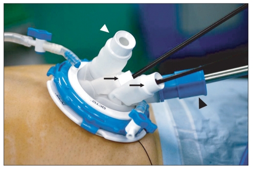
Purpose: Laparoscopic wedge resection of gastric submucosal tumor may be difficult in case of the endophytic mass or the mass located unreachable area such as cardia, and intragastric approach can be useful. We would present the experiences of the intragastric wedge resection.
Materials and methods: There were 7 patients diagnosed as gastric submucosal tumor and underwent the intragastric wedge resection at Surgery, Chungnam National University Hospital. We reviewed medical record.
Results: There were 3 male and 4 female. Mean age was 65 years-old (57~73). Mean body mass index was 26.28 kg/m(2) (21.28~35.30). Location of lesions was 4 cardia, 2 fundus and 1 midbody, respectively. Mean operation time was 83.6 minutes (70~105). All patients were healed without any complication. Mean postoperative hospital stay was 5.4 days (4~6). Mean size was 2.7 cm (2.3~3.8). Pathologic finding was 5 gastrointestinal stromal tumor and 2 leiomyoma.
Conclusions: The single incision intragastric wedge resection of gastric submucosal tumor is feasible and acceptable, especially in mass of gastric upper part.
Keywords: Gastrectomy; Gastrointestinal stromal tumors; Laparoscopy; Stomach neoplasms; Surgical procedures, minimally invasive.
Figures
References
-
- Kwon JG, Kim EY, Kim YS, Chun JW, Chung JT, You SS, et al. Accuracy of endoscopic ultrasonographic impression compared with pathologic diagnosis in gastrointestinal submucosal tumors. Korean J Gastroenterol. 2005;45:88–96. - PubMed
-
- Wiech T, Walch A, Werner M. Histopathological classification of nonneoplastic and neoplastic gastrointestinal submucosal lesions. Endoscopy. 2005;37:630–634. - PubMed
-
- Casali PG, Jost L, Reichardt P, Schlemmer M, Blay JY ESMO Guidelines Working Group. Gastrointestinal stromal tumours: ESMO clinical recommendations for diagnosis, treatment and follow-up. Ann Oncol. 2009;20(Suppl 4):64–67. - PubMed
-
- Ryu KJ, Jung SR, Choi JS, Jang YJ, Kim JH, Park SS, et al. Laparoscopic resection of small gastric submucosal tumors. Surg Endosc. 2011;25:271–277. - PubMed
LinkOut - more resources
Full Text Sources