. 2012 Feb 9;73(3):445-52.
doi: 10.1016/j.neuron.2011.11.026.

Differential effects of unfolded protein response pathways on axon injury-induced death of retinal ganglion cells

Affiliations

Differential effects of unfolded protein response pathways on axon injury-induced death of retinal ganglion cells

Yang Hu et al. Neuron. .

Erratum in

Abstract

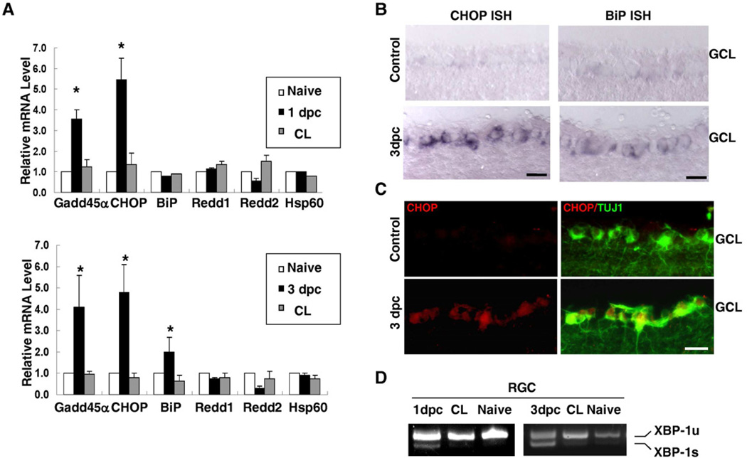
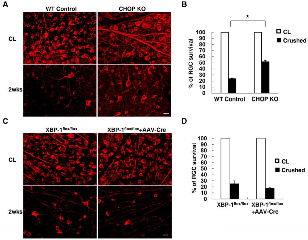
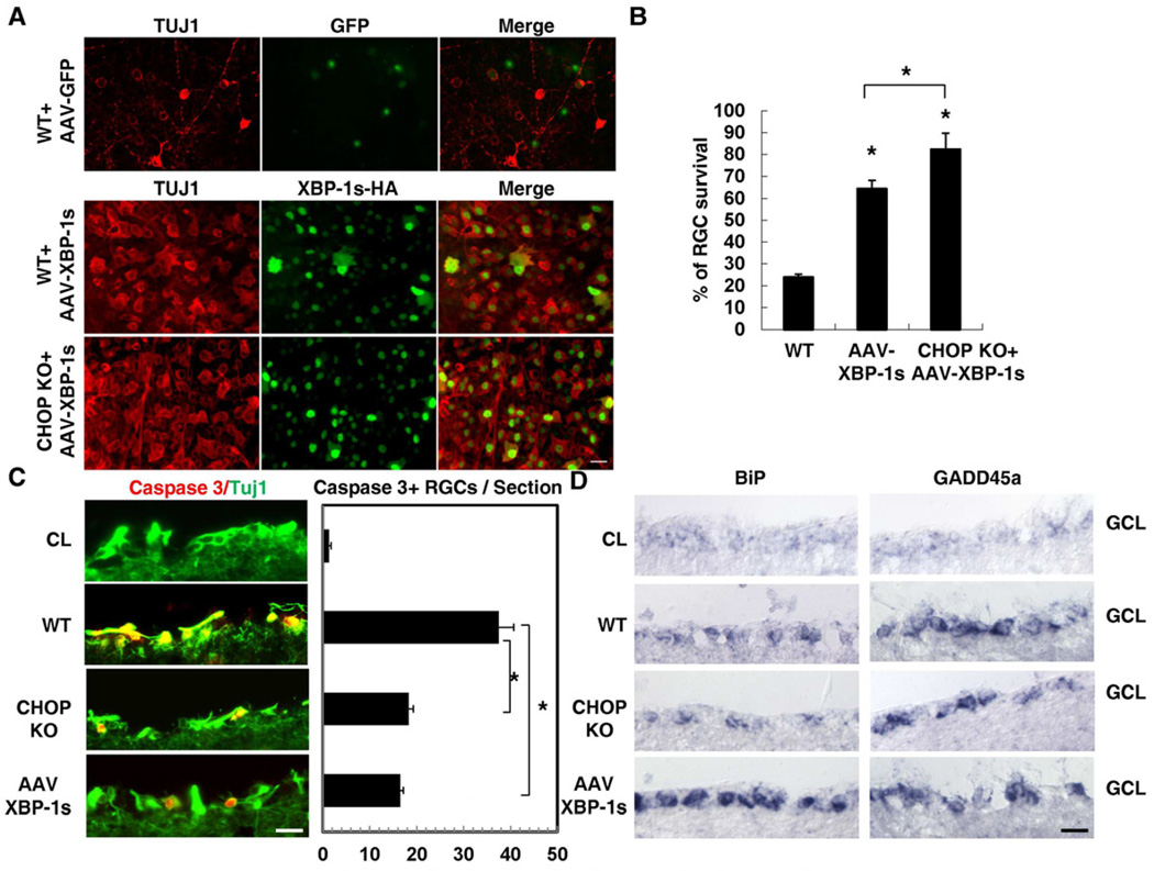
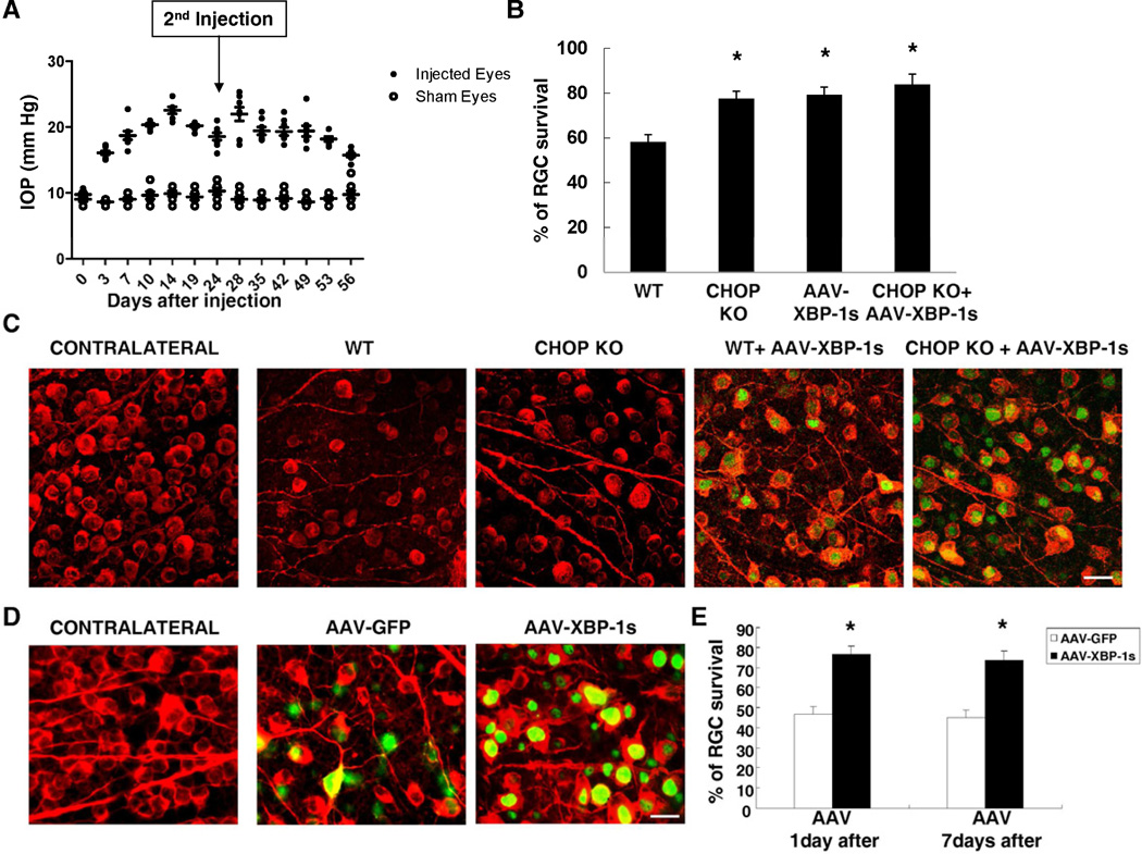
Loss of retinal ganglion cells (RGCs) accounts for visual function deficits after optic nerve injury, but how axonal insults lead to neuronal death remains elusive. By using an optic nerve crush model that results in the death of the majority of RGCs, we demonstrate that axotomy induces differential activation of distinct pathways of the unfolded protein response in axotomized RGCs. Optic nerve injury provokes a sustained CCAAT/enhancer binding homologous protein (CHOP) upregulation, and deletion of CHOP promotes RGC survival. In contrast, IRE/XBP-1 is only transiently activated, and forced XBP-1 activation dramatically protects RGCs from axon injury-induced death. Importantly, such differential activations of CHOP and XBP-1 and their distinct effects on neuronal cell death are also observed in RGCs with other types of axonal insults, such as vincristine treatment and intraocular pressure elevation, suggesting a new protective strategy for neurodegeneration associated with axonal damage.


Figures

Figure 1
Figure 1. Optic Nerve Injury Induces UPR in Adult RGCs
(A) Quantification of expression of different molecules in RGCs detected by q-PCR. Each sample was run in quadruplicate in each assay. GADPH was used as the endogenous control. CL: contralateral uninjured eye. *: p <0.01, paired Student’s t-test. Data are presented as means ± s.e.m and n=6. (B) In situ hybridization (ISH) results showing the expression of CHOP and BiP in the ganglion cell layer of adult mouse retinas. Scale bar: 20 µm. 3dpc: 3 days post-crush. GCL: ganglion cell layer. (C) Immunohistochemical analysis for CHOP or TUJ1 immunoreactivity in retinal sections. Scale bar: 20 µm. GCL: ganglion cell layer. (D) Detection of un-spliced and spliced XBP-1 mRNA (XBP-1u or XBP-1s) by RT-PCR. The mRNAs were prepared from retrograde-labeled and FACS-purified adult RGCs from retinas one day, three days post-crush (1dpc, 3dpc), contralateral (CL) eyes and naïve eyes. See also Figure S2.
Figure 2
Figure 2. CHOP KO, but not XBP-1 KO, Increases RGC survival after Optic Nerve Injury
Representative images (A, C) of fluorescent photomicrographs of flat-mounted retinas showing surviving TUJ1+ RGCs at 2 weeks after injury in WT mice and CHOP KO mice (A), and XBP-1flox/flox control mice (no AAV-Cre) and XBP-1flox/flox mice injected with AAV-Cre (C). CL: contralateral uninjured eye. Scale bar, 20 µm. (B, D) Quantification of surviving RGCs, represented as percentage of TUJ1+ RGCs, compared to the uninjured contralateral retinas, in CHOP KO mice (B) or XBP1 KO mice (D). *: p<0.01, Student’s t-test. Data are presented as means ± s.e.m and n = 6. See also Figure S1.
Figure 3
Figure 3. The Effects of XBP-1s Overexpression on RGC Survival after Optic Nerve Injury
(A) Representative images of flat-mounted retinas showing surviving TUJ1+ RGCs at 2 weeks post-injury in WT control mice injected with AAV-GFP or AAV-XBP-1s or CHOP KO mice injected with AAV-XBP-1s. Anti-HA antibody is used to indicate RGCs injected with AAVs expressing his-tagged XBP-1s. Scale bar, 20 µm (B) Quantification of TUJ1+ RGCs represented as percentage of TUJ1+ RGCs compared to the uninjured contralateral retinas. Data are presented as means ± s.e.m and n=6. * p<0.01, One way ANOVA and Tukey's Multiple Comparison Test. (C) Left panel: immunostaining of active caspase-3 at 3 days post-injury in WT, CHOP KO mice and AAV-XBP-1s injected mice. CL: uninjured contralateral eye. Scale bar: 20 µm. Right panel: quantification of caspase-3+ RGCs per section. Data are presented as means ± s.e.m and n=4. * p<0.01, One way ANOVA and Tukey's Multiple Comparison Test. (D) In situ hybridization results showing the expression of BiP and GADD45αin the ganglion cell layer (GCL) of WT, CHOP KO mice and AAV-XBP-1s injected mouse retinas 3 days post-injury. CL: contralateral uninjured eye. Scale bar: 20 µm. See also Figure S3.
Figure 4
Figure 4. CHOP Deletion and XBP-1s Over-expression Protect RGCs after IOP Elevation
(A) Measured IOP levels in the eyes of the wild type mice with saline injection (sham eyes) or microbeads injection (injected eyes). As the sham eyes exhibited a steady level of IOP at 10.0 ± 1.5 mmHg (n = 8), a single injection induced IOP elevation which lasts for 4 weeks and a second dose of microbeads injection on day 24 (arrow) could maintain elevated IOP levels to 8 weeks (n=8). (B) Quantification of surviving RGCs, represented as percentage of TUJ1+ RGCs, compared to the uninjured contralateral retinas, in each group at 8 weeks after IOP elevation. Data are presented as means ± s.e.m and n = 8. * p<0.01; One way ANOVA and Tukey's Multiple Comparison Test. (C) Representative images of TUJ1 (red) and HA (green) labeling in flat-mounted retinas from uninjected contralateral eyes, and from those eyes 8 weeks after IOP elevation. AAV-XBP-1s were injected two weeks before IOP elevation. Scale bar, 20 µm. (D) Representative images of TUJ1 (red) and GFP or HA (green) labeling in flat-mounted retinas from uninjected contralateral eyes, and from those eyes 8 weeks after IOP elevation. AAV-XBP-1s were injected 1 day after IOP elevation. Scale bar, 20 µm. (E) Quantification of surviving RGCs, represented as percentage of TUJ1+ RGCs, compared to the uninjured contralateral retinas, in each group at 8 weeks after IOP elevation. AAV-XBP-1s were injected 1 day or 7 days after IOP elevation. Data are presented as means ± s.e.m and n = 8. * p<0.01; Student’s t-test. See also Figure S4.

Comment in

References

    1. Aoki S, Su Q, Li H, Nishikawa K, Ayukawa K, Hara Y, Namikawa K, Kiryu-Seo S, Kiyama H, Wada K. Identification of an axotomy-induced glycosylated protein, AIGP1, possibly involved in cell death triggered by endoplasmic reticulum-Golgi stress. J Neurosci. 2002;22:10751–10760. - PMC - PubMed
    1. Berkelaar M, Clarke DB, Wang YC, Bray GM, Aguayo AJ. Axotomy results in delayed death and apoptosis of retinal ganglion cells in adult rats. J Neurosci. 1994;14:4368–4374. - PMC - PubMed
    1. Brugarolas J, Lei K, Hurley RL, Manning BD, Reiling JH, Hafen E, Witters LA, Ellisen LW, Kaelin WG., Jr Regulation of mTOR function in response to hypoxia by REDD1 and the TSC1/TSC2 tumor suppressor complex. Genes Dev. 2004;18:2893–2904. - PMC - PubMed
    1. Busskamp V, Duebel J, Balya D, Fradot M, Viney TJ, Siegert S, Groner AC, Cabuy E, Forster V, Seeliger M, et al. Genetic reactivation of cone photoreceptors restores visual responses in retinitis pigmentosa. Science. 2010;329:413–417. - PubMed
    1. Calfon M, Zeng H, Urano F, Till JH, Hubbard SR, Harding HP, Clark SG, Ron D. IRE1 couples endoplasmic reticulum load to secretory capacity by processing the XBP-1 mRNA. Nature. 2002;415:92–96. - PubMed

Publication types

MeSH terms