. 2012 Feb 9;73(3):482-96.
doi: 10.1016/j.neuron.2011.11.021.

Palmitoylation by DHHC5/8 targets GRIP1 to dendritic endosomes to regulate AMPA-R trafficking

Affiliations

Palmitoylation by DHHC5/8 targets GRIP1 to dendritic endosomes to regulate AMPA-R trafficking

Gareth M Thomas et al. Neuron. .

Abstract

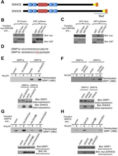
Palmitoylation, a key regulatory mechanism controlling protein targeting, is catalyzed by DHHC-family palmitoyl acyltransferases (PATs). Impaired PAT activity is linked to neurodevelopmental and neuropsychiatric disorders, suggesting critical roles for palmitoylation in neuronal function. However, few substrates for specific PATs are known, and functional consequences of palmitoylation events are frequently uncharacterized. Here, we identify the closely related PATs DHHC5 and DHHC8 as specific regulators of the PDZ domain protein GRIP1b. Binding, palmitoylation, and dendritic targeting of GRIP1b require a PDZ ligand unique to DHHC5/8. Palmitoylated GRIP1b is targeted to trafficking endosomes and may link endosomes to kinesin motors. Consistent with this trafficking role, GRIP1b's palmitoylation turnover rate approaches the highest of all reported proteins, and palmitoylation increases GRIP1b's ability to accelerate AMPA-R recycling. To our knowledge, these findings identify the first neuronal DHHC5/8 substrate, define novel mechanisms controlling palmitoylation specificity, and suggest further links between dysregulated palmitoylation and neuropathological conditions.


Figures

Figure 1
Figure 1. DHHC5 and DHHC8 specifically bind and palmitoylate GRIP1
A: Schematic of the structure of DHHC5 and DHHC8, showing predicted transmembrane domains (blue), catalytic DHHC-Cysteine Rich Domain (red) and identical C-terminal 15 AA sequence (yellow) terminating in identical PDZ ligand (EISV: orange). The region of the DHHC8 C-terminus used as bait for yeast two-hybrid screening is indicated. B: HEK293T cells were transfected with myc-tagged GRIP1-PDZ domains 4 thru 6, together with either GST alone (GST), a GST fusion of the C-terminus of DHHC5 (GST-5wt), or a GST fusion of the C-terminus of DHHC5 lacking the C-terminal PDZ-binding motif (GST-5ΔC). Inputs (left panels) and GST pulldowns (right panels) were immunoblotted with anti-myc and anti-GST antibodies. C: same as B, except that indicated constructs of DHHC8 were cotransfected. D: Comparison of N-terminal sequences of GRIP1a and GRIP1b. The unique Cysteine11 of GRIP1b is reported to be palmitoylated in heterologous cells, but the PAT was not identified. E: HEK293T cells were transfected with HA-tagged DHHC5 and either full-length untagged GRIP1a or GRIP1b. Acyl-biotin exchange (ABE) was performed to isolate palmitoylated proteins and GRIP1 levels in ABE samples were detected by immunoblotting (top panel). Cell lysates were blotted to detect total levels of GRIP1 (middle panel) and HA-DHHC5 (lower panel). Note that palmitoylated GRIP1 signal is only detected following treatment with hydroxylamine (NH2OH), an essential step of the ABE reaction. F: as E, except that cells were transfected with mycHis-DHHC8 and either GRIP1a or GRIP1b and lysates were blotted to detect total levels of GRIP1 (middle panel) and mycHis-DHHC8 (lower panel). G: Palmitoylation of GRIP1b by DHHC5 requires catalytic activity and PDZ binding ability. HEK293T cells were transfected with GRIP1b plus either empty vector, HA-tagged DHHC5 wild-type (HA-DHHC5wt), catalytically inactive mutant (HA-DHHS5) or PDZ ligand mutant (HA-DHHC5ΔC). Lysates were subjected to acyl-biotinyl exchange (ABE) and immunoblotted to detect palmitoylated GRIP1 (upper panel), GRIP1 expression (middle panel) and DHHC5 expression (lower panel). H: as G, except that cells were transfected with GRIP1b plus either DHHC8wt, DHHS8 or DHHC8ΔC as indicated. See also Supplemental Figure S1 for additional control experiments and quantified data.
Figure 2
Figure 2. DHHC5/8 control GRIP1 palmitoylation in primary neurons
A: Top row panels: Primary hippocampal neurons were immunostained with antibodies against endogenous DHHC5 (first panel), DHHC8 (second panel) and the synaptic marker Bassoon (third panel). Lower panels show high magnification images of a single highlighted dendrite. Examples of colocalized DHHC8 and Bassoon puncta are indicated with arrows in the overlaid image. A scale bar (20 μm) is marked in this and subsequent images. B: Immunostaining of primary hippocampal neurons cotransfected with myc-tagged DHHC5 and the soluble marker mCherry, and immunostained for the synaptic marker PSD-95 plus myc and dsRed antibodies. Lower panels show high magnification images of dendrites. An example of myc-DHHC5 colocalized with a PSD-95-positive dendritic spine is shown (arrow) but myc-DHHC5 is more frequently found in dendritic shafts. C: Equal protein amounts of the indicated subcellular fractions were immunoblotted with DHHC5 or DHHC8 antibodies. Fidelity of the preparation was confirmed by immunoblotting with PSD-95 antibody. D: Dendritic GRIP1 puncta frequently contain the GRIP1b isoform. Hippocampal neurons were immunostained with GRIP1 monoclonal and GRIP1b-specific polyclonal antibodies as indicated. Lower panels show high magnification images of dendrites. GRIP1 dendritic puncta that are GRIP1b-positive are indicated with arrows in the overlaid image. E: High stoichiometry of GRIP1b palmitoylation in primary neurons and whole brain. Top panels: Cultured neurons metabolically labeled with [3H]-palmitate were lysed and immunoprecipitated with the indicated antibodies. [3H] signal (upper panel) and GRIP1 protein levels (lower panel) are shown. Bottom panels: Cultured neurons were subjected to ABE and immunoblotted with pan-GRIP1 (upper panel) or GRIP1b-specific antibodies (lower panel). F: Lysates from cortical neurons cultured for the indicated number of days in vitro (DIV) were immunoblotted for expression of GRIP1b (top panel) or DHHC5 (second panel). Lysates subjected to ABE were blotted to detect palmitoylated GRIP1b (bottom panel). G: Cortical cultured neurons were infected with the indicated lentiviruses containing shRNA sequences to knock down expression of DHHC5, DHHC8 or both PATs. A fraction of each lysate was immunoblotted to detect total levels of DHHC5 (top panel), GRIP1 (third panel) and Fyn (fifth panel). The remaining lysate was subjected to ABE to detect palmitoylated GRIP1 (second panel) and Fyn (fourth panel). H: Quantitation of multiple experiments confirms that DHHC5 knockdown significantly reduces GRIP1 palmitoylation (56.8 ± 7.2% of control virus, n=8, p<0.05 compared to control virus (FUGW) alone, t-test), shRNA-resistant DHHC5 (`rescue') restores GRIP1 palmitoylation levels (95.5 ± 7.5% of control virus, n=8, not significant from FUGW alone) and GRIP1 palmitoylation is almost completely eliminated by knockdown of both DHHC5 and DHHC8 (6.9 ± 2.6% of control virus, n=3, p<0.05 compared to FUGW alone, t-test). See also Supplemental Figure S2.
Figure 3
Figure 3. Palmitoylation targets GRIP1b to specific dendritic endosomal vesicles
A: Primary neurons were treated with 100 μM 2-Bromopalmitate for the indicated times or with 0.1% (v/v) EtOH (solvent control). ABE reactions were performed to detect palmitoylated and total levels of GRIP1 and PSD-95, as indicated. B: Quantitation of palmitate turnover from multiple experiments as in A, plotted as Mean ± SEM for n=4 determinations per time point. C: Schematic of palmitoylated GRIP1b wild type, compared to non-palmitoylatable (GRIP1b-C11S) and constitutive palmitolyation-mimic (G1b-Myr). The Myristoylation consensus adds a similar lipid (C14, saturated) at amino acid 2 (Gly), following cleavage of the initiating Methionine. DF: Dendritic targeting of GRIP1b by lipid modification. Representative images of primary hippocampal neurons transfected with GFP as a morphology marker, plus either GRIP1bwt-myc (GRIP1wt, top row), GRIP1b-C11S-myc (GRIP1CS, middle row) or myristoylated GRIP1b-myc (Myr-GRIP1, bottom row), following fixation and staining with antibodies against GFP (first column) and myc (second column). The right column shows myc signals, thresholded at an identical value for each transfected neuron. E: Quantitation of average fluorescence in cell soma and in dendritic segments at the indicated distances from the soma, in neurons expressing GRIP1bwt-myc (n=14), GRIP1b-C11S-myc (n=16) or myristoylated GRIP1b-myc (n=15). Asterisks indicate significant difference (p<0.05) from GRIP1bwt. F: Number of GRIP1b puncta per dendritic segment for each neuron from E (plotted for 3 dendrites each from n=14, n=16 and n=15 neurons) at the indicated distances from the cell soma. G: Mimicking palmitoylation targets GRIP1b to recycling endosomes. Myr-GRIP1b-myc transfected neurons were live labeled with Alexa555-transferrin, fixed and stained with anti-myc antibodies. Lower panels show high magnification images of an individual dendrite. Extensively colocalized dendritic puncta are indicated with white arrows in the overlaid image. See also Supplemental Figure S3.
Figure 4
Figure 4. Catalytic activity and PDZ domain recognition by DHHC5 target GRIP1bwt to dendritic vesicles
A: Representative images of primary hippocampal neurons transfected with GFP as a morphology marker, GRIP1bwt-myc or GRIP1b-C11S-myc and either empty vector, HA-DHHC5wt, HA-DHHS5 or DHHC5deltaC, following fixation and staining with antibodies against GFP (first column), myc (second column) and HA tags (fourth panels). The third column shows myc signal, thresholded at an identical value for each transfected neuron. B: Average fluorescence intensity of GRIP1b-myc was plotted as in Fig 3E for GRIP1bwt-myc (n=14), GRIP1bwt-myc plus HA-DHHC5wt (n=19), GRIP1bC11S plus HA-DHHC5wt (n=12), GRIP1bwt plus HA-DHHS5 (n=15) and GRIP1bwt plus HA-DHHC5ΔC (n=14). C: Dendritic puncta of GRIP1b-myc were counted as in Fig 3F. Data for GRIP1bwt-myc from Figure 3 are re-plotted in panels 4B and 4C. Asterisks indicate significant difference (P<0.05, t-test) from GRIP1bwt alone. N.B. Quantitative analysis of DHHC5 dendritic distribution confirmed that DHHC5 mutants target equally well, if not better than, DHHC5wt to distal dendrites (See also Supplemental Figure S4), thus impaired targeting does not account for their inability to regulate GRIP1bwt distribution.
Figure 5
Figure 5. Rapid turnover of palmitate on GRIP1 modulates dendritic targeting
A: Acute treatment with 2-Bromopalmitate disperses dendritic GRIP1 puncta. Top panels: Unprocessed images of GRIP1 immunostaining in GFP-transfected hippocampal neurons. Middle panels: GFP signal (morphology marker to outline individual dendrites). Lower panels: Thresholded puncta within the boundary of an individual GFP-transfected neuron following treatment with EtOH vehicle or 100 μM 2-Bromopalmitate for 90 minutes. Right panel: Plot of number of endogenous GRIP1 puncta (mean ± SEM) counted at indicated distances from the cell soma for 3 dendrites per cell from n=19 cells (control) and n=21 cells (2-Br). B: Membrane-associated GRIP1b preferentially binds dendritic kinesin KIF5C in heterologous cells. HEK293 cells were transfected with HA-tagged KIF5C plus the indicated myc-tagged GRIP1 constructs. Myc immunoprecipitates were immunoblotted for co-immunoprecipitated HA-KIF5C. C: Deletion of Kinesin-binding-domain (KBD) impairs Myr-GRIP1b dendritic targeting. Top panels: representative images of primary hippocampal neurons transfected with GFP as a morphology marker, plus either full length myristoylated GRIP1b-myc (Myr-GRIP1b) or Myr-GRIP1b lacking KBD (Myr-GRIP1b-delKBD), following fixation and staining with antibodies against GFP (first row) and myc (second row). The bottom row shows myc signals, thresholded at an identical value for each transfected neuron. D: Average fluorescence intensity of myc-tagged constructs in soma and dendrites, plotted as in Fig 3E for Myr-GRIP1b (n=13) and Myr-GRIP1b-delKBD (n=12). Double Asterisks indicate significant differences (p<0.01, t-test) between conditions at all distances from soma.
Figure 6
Figure 6. Targeting GRIP1b to membranes increases activity-dependent GluA2 recycling
A: Representative images of fluorescence change of a hippocampal neuron expressing pH-GluA2, exposed at t=10 to 20 μM NMDA for 5 min and then allowed to recover following NMDA washout. B: Fluorescence change (mean ± s.e.m.) during initial incubation, perfusion at t= 10 min with 20 μM NMDA for 5 min (blue bar) and washout, plotted for hippocampal neurons transfected with pHluorin-tagged GluA2 (pHGluA2) plus either empty vector (black circles, n=11), myc-tagged GRIP1b wildtype (G1bwt, red circles, n=5) or GRIP1b-C11S (G1bCS, blue circles, n=7). C, D: As B, but for neurons transfected with pH-GluA2 plus myristoylated GRIP1b (Myr-G1b, orange circles, n=6), or with HA-tagged DHHC5 (green circles, n=8), respectively. Recycling in DHHC5-transfected neurons is still accelerated when the fluorescence decrease is scaled to match vector controls (Fig S5A). E: Time constants (T1/2) of fluorescence recovery, plotted as mean ± SEM for the curves in B–D. Asterisks indicate significant difference from pHGluA2 alone (P<0.05, t test). See also Supplemental Figure S5.

References

    1. Ashby MC, De La Rue SA, Ralph GS, Uney J, Collingridge GL, Henley JM. Removal of AMPA receptors (AMPARs) from synapses is preceded by transient endocytosis of extrasynaptic AMPARs. J. Neurosci. 2004;24:5172–5176. - PMC - PubMed
    1. Chen WY, Shi YY, Zheng YL, Zhao XZ, Zhang GJ, Chen SQ, Yang PD, He L. Case-control study and transmission disequilibrium test provide consistent evidence for association between schizophrenia and genetic variation in the 22q11 gene ZDHHC8. Hum. Mol. Genet. 2005;13:2991–2995. - PubMed
    1. Daw MI, Chittajallu R, Bortolotto ZA, Dev KK, Duprat F, Henley JM, Collingridge GL, Isaac JT. PDZ proteins interacting with C-terminal GluR2/3 are involved in a PKC-dependent regulation of AMPA receptors at hippocampal synapses. Neuron. 2000;28:873–876. - PubMed
    1. DeSouza S, Fu J, States BA, Ziff EB. Differential palmitoylation directs the AMPA receptor-binding protein ABP to spines or to intracellular clusters. J. Neurosci. 2002;22:3493–3503. - PMC - PubMed
    1. Dong H, O'Brien RJ, Fung ET, Lanahan AA, Worley PF, Huganir RL. GRIP: a synaptic PDZ domain-containing protein that interacts with AMPA receptors. Nature. 1997;386:279–284. - PubMed

Publication types

MeSH terms