. 2012 Feb 8;102(3):523-31.
doi: 10.1016/j.bpj.2011.12.049. Epub 2012 Feb 7.

The role of lipids in VDAC oligomerization

Affiliations

The role of lipids in VDAC oligomerization

Viktoria Betaneli et al. Biophys J. .

Abstract

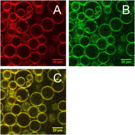
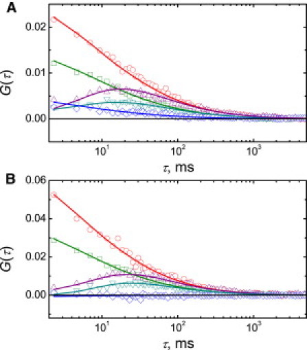
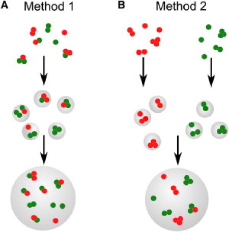
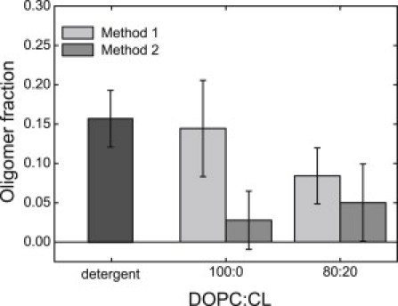
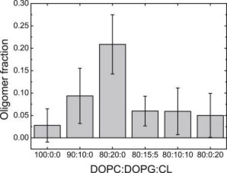
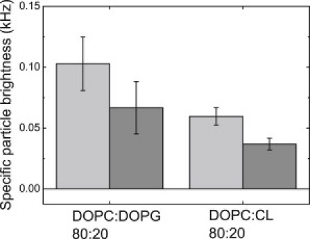
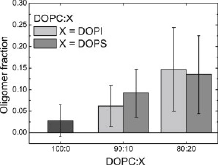
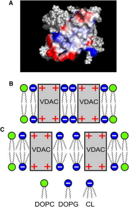
Evidence has accumulated that the voltage-dependent anion channel (VDAC), located on the outer membrane of mitochondria, plays a central role in apoptosis. The involvement of VDAC oligomerization in apoptosis has been suggested in various studies. However, it still remains unknown how exactly VDAC supramolecular assembly can be regulated in the membrane. This study addresses the role of lipids in this process. We investigate the effect of cardiolipin (CL) and phosphatidylglycerol (PG), anionic lipids important for mitochondria metabolism and apoptosis, on VDAC oligomerization. By applying fluorescence cross-correlation spectroscopy to VDAC reconstituted into giant unilamellar vesicles, we demonstrate that PG significantly enhances VDAC oligomerization in the membrane, whereas cardiolipin disrupts VDAC supramolecular assemblies. During apoptosis, the level of PG in mitochondria increases, whereas the CL level decreases. We suggest that the specific lipid composition of the outer mitochondrial membrane might be of crucial relevance and, thus, a potential cue for regulating the oligomeric state of VDAC.


Figures

Figure 1
Figure 1
Representative confocal fluorescence microscopy images of GUVs with reconstituted red- and green-labeled VDAC: (A) red (ATTO 655) channel, (B) green (Alexa 488) channel, and (C) merge of panels A and B. GUV composition: DOPC/DOPG/CL 75:15:5.
Figure 2
Figure 2
Representative scanning FCCS auto- and cross-correlation curves of fluorescence intensity fluctuations of red- and green-labeled VDAC reconstituted into GUVs. Experimental FCS data (symbols) are shown along with the corresponding least-squares fits: single-focus, single-color, red-channel autocorrelation (circles); single-focus, single-color, green-channel autocorrelation (squares); two-focus, single-color, red channel cross-correlation (up-triangles); two-focus, single-color, green-channel cross-correlation (down-triangles); and one-focus, two-color, red-green cross-correlation (diamonds). GUV composition: (A) DOPC/DOPG 80:20, (B) DOPC/CL 80:20. Temperature, 21 ± 0.5°C.
Figure 3
Figure 3
Two different methods of reconstitution of fluorescently labeled VDAC into GUVs. (A) VDACred and VDACgreen are mixed in detergent solution and incorporated into proteoliposomes, which are further used to form GUVs. (B) VDACred and VDACgreen are incorporated into proteoliposomes separately, and the proteoliposomes are mixed only immediately before the formation of GUVs.
Figure 4
Figure 4
Oligomer fraction of fluorescently labeled VDAC in detergent solution (dark shaded) and upon reconstitution into GUVs with two lipid compositions: pure DOPC and DOPC/CL 80:20. VDACred and VDACgreen were incorporated into GUVs using the two different methods shown in Fig. 3: Method 1 (light shaded) or Method 2 (medium shaded). Data were obtained in three independent experiments with three independently prepared samples; each point is an average of values measured on 10–40 vesicles.
Figure 5
Figure 5
Oligomer fraction of fluorescently labeled VDAC in GUVs. GUV composition: DOPC, DOPC/DOPG 90:10, DOPC/DOPG 80:20, DOPC/DOPG/CL 80:15:5, DOPC/DOPG/CL 80:10:10, and DOPC/CL 80:20. Data were obtained in three independent experiments with three independently prepared samples; each point is an average of values measured on 13–39 vesicles.
Figure 6
Figure 6
Specific particle brightness of fluorescently labeled VDACgreen in GUVs. GUV composition: DOPC/DOPG 80:20 or DOPC/CL 80:20. (Light-shaded and medium-shaded) Data depict two independent experiments carried out with two independently prepared samples. Each point is an average of values measured on 5–7 vesicles.
Figure 7
Figure 7
Oligomer fraction of fluorescently labeled VDAC in GUVs. GUV composition: DOPC (dark shaded), DOPC/DOPI 90:10, DOPC/DOPI 80:20 (light shaded) or DOPC/DOPS 90:10, and DOPC/DOPS 80:20 (medium shaded). Data were obtained in three independent experiments with three independently prepared samples; each point is an average of values measured on 13–29 vesicles.
Figure 8
Figure 8
Charge-surface (positive, red, and negative, blue) of VDAC monomer responsible for the dimer formation generated using the software PyMOL 1.3 (DeLano Scientific) (A) and schematic representation of the interaction of VDAC with anionic lipids (B and C). (B) Formation of VDAC dimers in presence of PG. (C) Interaction of VDAC with CL.

References

    1. Colombini M. VDAC: the channel at the interface between mitochondria and the cytosol. Mol. Cell. Biochem. 2004;256-257:107–115. - PubMed
    1. Tajeddine N., Galluzzi L., Kroemer G. Hierarchical involvement of Bak, VDAC1 and Bax in cisplatin-induced cell death. Oncogene. 2008;27:4221–4232. - PubMed
    1. Madesh M., Hajnóczky G. VDAC-dependent permeabilization of the outer mitochondrial membrane by superoxide induces rapid and massive cytochrome c release. J. Cell Biol. 2001;155:1003–1015. - PMC - PubMed
    1. Shimizu S., Matsuoka Y., Tsujimoto Y. Essential role of voltage-dependent anion channel in various forms of apoptosis in mammalian cells. J. Cell Biol. 2001;152:237–250. - PMC - PubMed
    1. Keinan N., Tyomkin D., Shoshan-Barmatz V. Oligomerization of the mitochondrial protein voltage-dependent anion channel is coupled to the induction of apoptosis. Mol. Cell. Biol. 2010;30:5698–5709. - PMC - PubMed

Publication types

LinkOut - more resources