. 2012 May;119(5):956-64.
doi: 10.1016/j.ophtha.2011.10.036. Epub 2012 Feb 9.

Mapping the nerve architecture of diabetic human corneas

Affiliations

Mapping the nerve architecture of diabetic human corneas

Jiucheng He et al. Ophthalmology. 2012 May.

Abstract

Objective: To investigate the entire human corneal nerve architecture of donors with different durations of insulin-dependent diabetes mellitus (IDDM).

Design: Experimental study.

Participants and controls: Sixteen fresh human eyes from 8 diabetic donors (aged 43-66 years, with IDDM for 2-17 years) and 12 eyes from 6 normal donors (aged from 44-67 years) were obtained from the National Disease Research Interchange (NDRI).

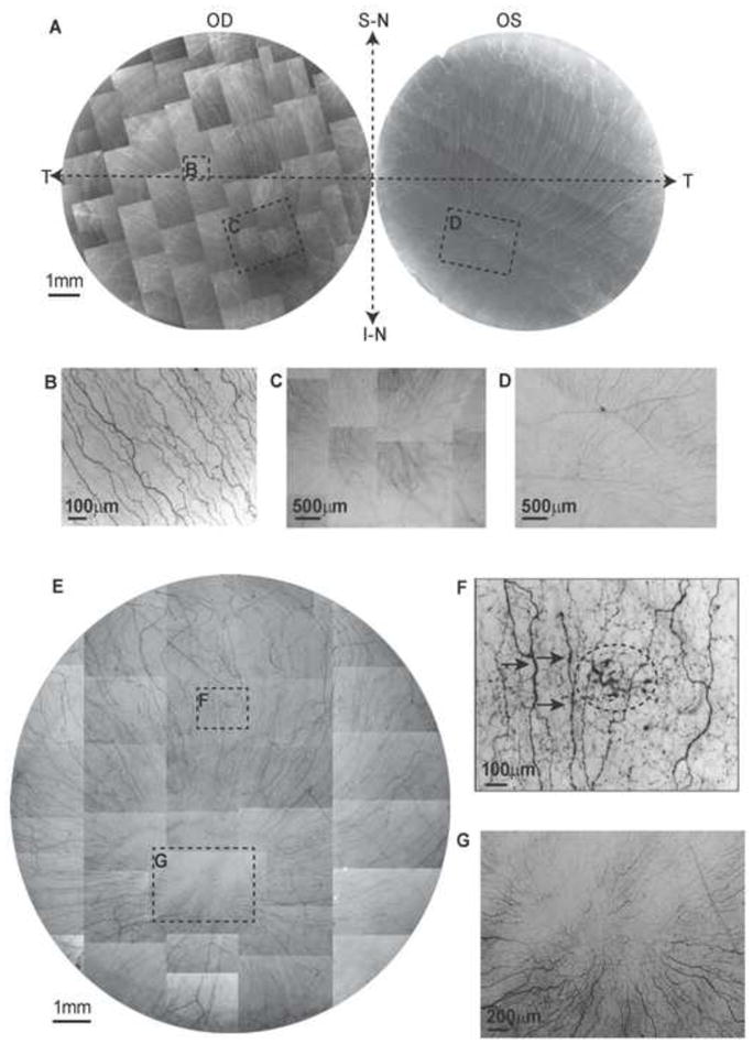
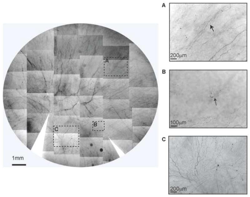
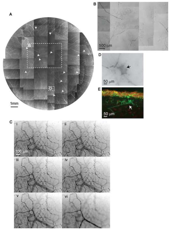
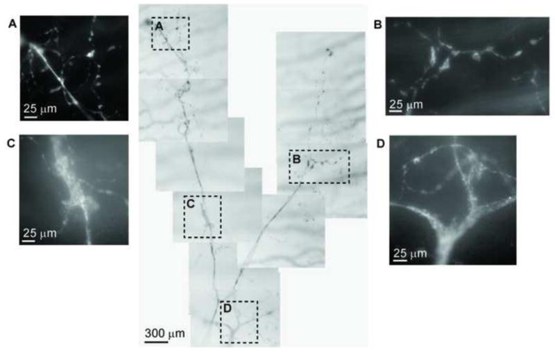
Methods: After fixation, corneas were stained with mouse monoclonal anti-β-Tubulin III antibody, and images were acquired to build a whole view of the corneal nerve architecture. The same corneas were used for both whole-mount and cross-section examination.

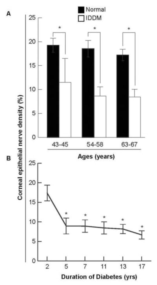
Main outcome measures: Corneal epithelial nerve density was calculated on the basis of the whole-mount view of the central area. The number of stromal nerves was calculated by counting the nerve trunks at the corneoscleral limbus of the entire cornea. Differences between diabetic and normal corneas in epithelial nerve densities and main stromal nerve numbers were compared by paired-samples t test.

Results: The diabetic eyes presented numerous neuropathies in areas where the epithelial nerve bundles emerged. A striking pathologic change was the presence of abundant nerve fiber loops in the stroma. These loops seemed to form by resistance presented by the basement membrane, which may prevent penetration of stromal nerve branches into epithelia. There was no difference in the numbers of main stromal nerve trunks between corneas from diabetic and normal donors, but there was a significant decrease in central epithelial nerve density in the diabetic corneas. We did not find an age effect on this decrease. Instead, it was significantly affected by 5 or more years of IDDM.

Conclusions: This is the first study to show an entire view of the nerve architecture in human diabetic corneas. The decreased epithelial nerve density may result from the abnormalities of stromal nerve architecture and is affected by 5 or more years of IDDM. Although compensation for some nerve regeneration takes place, the alterations in the stromal nerves can explain the poor healing and persistent epithelial defects seen in diabetic patients.


Figures

Figure 1
Figure 1
Whole mount view of corneal epithelial nerve distribution in normal and diabetic corneas at early stage of the disease. A) Images were acquired in a time-lapse mode with a Nikon Eclipse TE200 and with a 5X lens in compliance with the natural shape of the cornea from the two eyes of a 64-year-old normal male donor. OD: oculus dexter (right eye); OS: oculus sinister (left eye); S-N: superior nasal quadrant; I-N: inferior nasal quadrant; T: temporal quadrant. The epithelial nerve bundles ran in a radial pattern from the periphery to merge in an area within the inferior-nasal quadrant beyond the corneal apex. Highlighted images showed the detailed architecture of the epithelial nerve distribution in the central (B), and merging areas (C, D). E) Images were taken from the right eye of a 45-year-old male with insulin-dependent diabetes mellitus (IDDM) for two years. Highlighted images show the detailed nerve distribution in the converging area (G). F) Image F, outlined with the small frame, shows the increased thickness in parts of the nerves (arrows) and a sub-epithelial neuropathy (circle).
Figure 2
Figure 2
Whole mount view of corneal epithelial nerve distribution in a diabetic cornea after prolonged insulin-dependent diabetes mellitus (IDDM). Images were taken from the left eye of a 58-year-old female with IDDM for 13 years. The montage of images outlined a frame that shows the nerve distribution in detail. The images (A, B) show newly-regenerating epithelial nerve leashes (arrows) with abundant empty spaces without innervations. Image C shows the detailed nerve distribution in the merging area. The long bundles running from the superior quadrants converge in a counter-clock pattern at the inferior quadrant close to the limbal area. Several short nerve bundles, representing newly regenerating nerves, are visible in the lower part.
Figure 3
Figure 3
A) Whole mount view of corneal stromal nerve distribution in a diabetic cornea taken from a 43-year-old male with insulin-dependent diabetes mellitus (IDDM) for 5 years. Numerous nerve fiber loops and neuropathic lesions (arrows) are present in the whole cornea. B) Highlighted image shows the nerve fiber loops present in the center. C) Topographic images of the same area show the detailed architecture of corneal nerve loops at different depths. Images were recorded in time-lapse mode from the stroma surface (I) to the middle of the stroma (VI). D) and E) are representative images of diabetic neuropathies in a whole mount and in a transected view.
Figure 5
Figure 5
A) Age-paired comparative analysis of corneal epithelial nerve density between normal and diabetic human eyes. The corneal epithelial nerve density was significantly decreased in the diabetic eyes when compared to the normal. Data are expressed as average percent ± standard deviation (SD) of 64 images from 4 corneas in the normal groups and 64–96 images from 4–6 corneas in the diabetic groups (* p< 0.01). B) The effect of insulin-dependent diabetes mellitus (IDDM) duration on corneal epithelial nerve density. A significant decrease (*p < 0.01) was observed in the corneas of patients diagnosed with IDDM for 5 years or more. Data expressed as average percent ± SD, with each time point having 32 images analyzed from two eyes except for 5 years and 7 years time points, both of which have 64 images analyzed from 4 eyes.
Figure 7
Figure 7
Corneal stromal nerve degeneration. The images reveal the montage of a main stromal nerve trunk together with its branches of a 56-year-old male donor with insulin-dependent diabetes mellitus (IDDM) for 7 years. Highlighted images show the details of nerve degeneration at different parts of the nerve.

References

    1. He J, Bazan NG, Bazan HE. Mapping the entire human corneal nerve architecture. Exp Eye Res. 2010;91:513–23. - PMC - PubMed
    1. Müller LJ, Marfurt CF, Kruse F, Tervo TM. Corneal nerves: structure, content and function. Exp Eye Res. 2003;76:521–42. - PubMed
    1. Riaz SS, Tomlinson DR. Neurotrophic factors in peripheral neuropathies: pharmacological strategies. Prog Neurobiol. 1996;49:125–43. - PubMed
    1. Kingsley RE, Marfurt CF. Topical substance P and corneal epithelial wound closure in the rabbit. Invest Ophthalmol Vis Sci. 1997;38:388–95. - PubMed
    1. Jones MA, Marfurt CF. Peptidergic innervation of the rat cornea. Exp Eye Res. 1998;66:421–35. - PubMed

Publication types