

Validating imaging biomarkers of cerebral edema in patients with severe ischemic stroke
- PMID: 22325573
- PMCID: PMC3529850
- DOI: 10.1016/j.jstrokecerebrovasdis.2012.01.002
Validating imaging biomarkers of cerebral edema in patients with severe ischemic stroke
Abstract
Background: There is no validated neuroimaging marker for quantifying brain edema. We sought to test whether magnetic resonance imaging (MRI)-based metrics would reliably change during the early subacute period in a manner consistent with edema and whether they would correlate with relevant clinical endpoints.
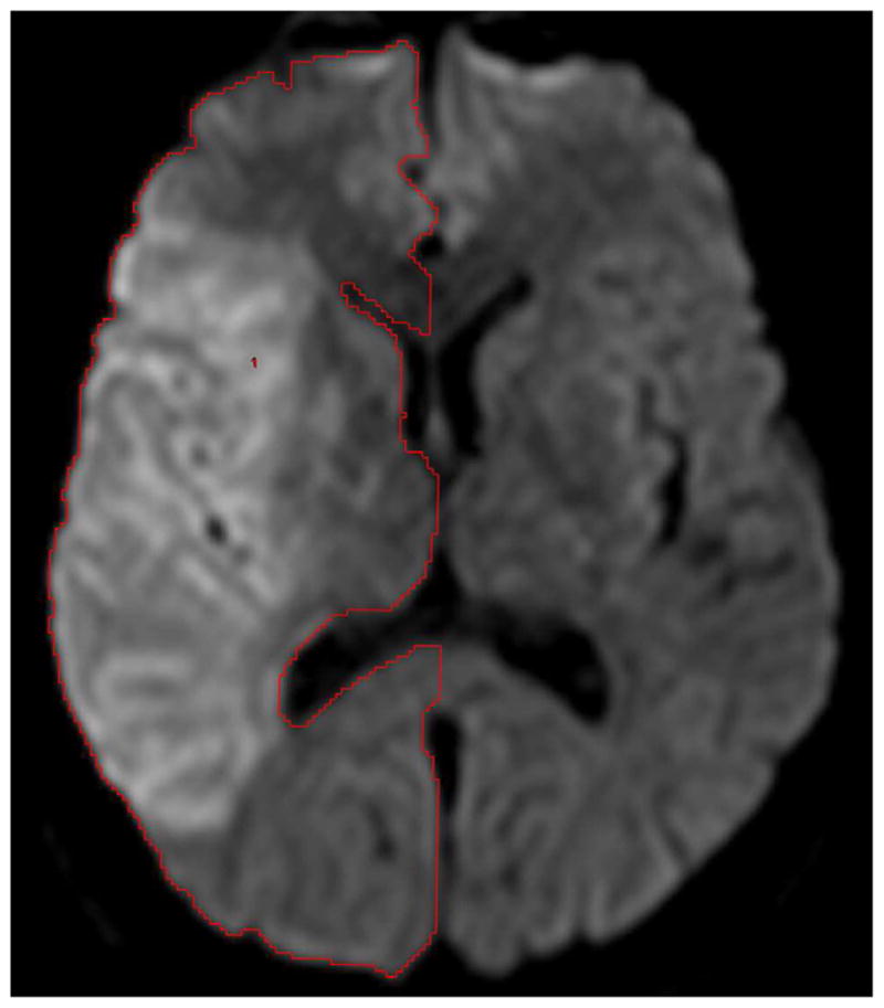
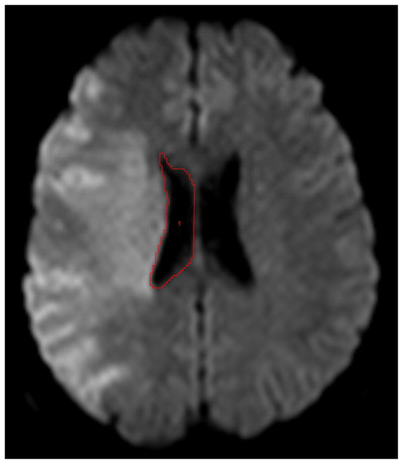
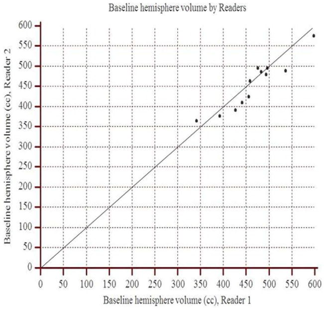
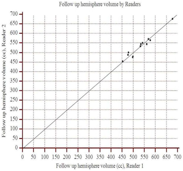
Methods: Serial MRI studies from patients in the Echoplanar Imaging Thrombolytic Evaluation Trial with initial diffusion-weighted imaging (DWI) lesion volume >82 cm(3) were analyzed. Two independent readers outlined the hemisphere and lateral ventricle on the involved side and calculated respective volumes at baseline and days 3 to 5. We assessed interrater agreement, volume change between scans, and the association of volume change with early neurologic deterioration (National Institutes of Health Stroke Scale score worsening of ≥ 4 points), a 90-day modified Rankin scale (mRS) score of 0 to 4, and mortality.
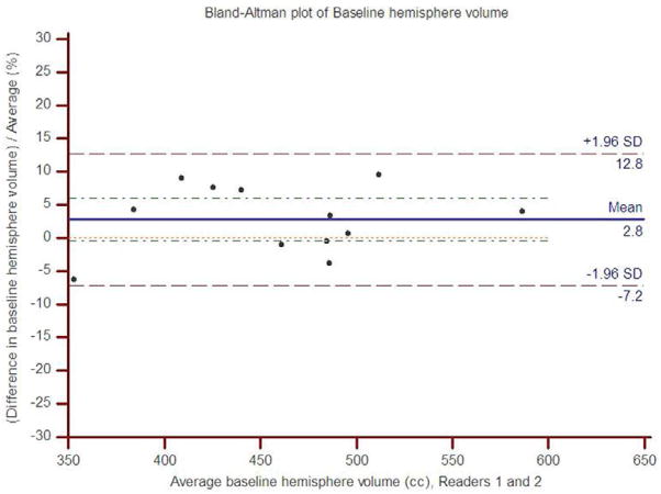
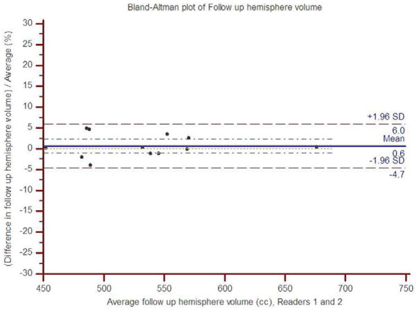
Results: Of 12 patients who met study criteria, average baseline and follow-up DWI lesion size was 138 cm(3) and 234 cm(3), respectively. The mean time to follow-up MRI was 62 hours. Concordance correlation coefficients between readers were >0.90 for both hemisphere and ventricle volume assessment. Mean percent hemisphere volume increase was 16.2 ± 8.3% (P < .0001), and the mean percent ventricle volume decrease was 45.6 ± 16.9% (P < .001). Percent hemisphere growth predicted early neurologic deterioration (area under the curve [AUC] 0.92; P = .0005) and 90-day mRS 0 to 4 (AUC 0.80; P = .02).
Conclusions: In this exploratory analysis of severe ischemic stroke patients, statistically significant changes in hemisphere and ventricular volumes within the first week are consistent with expected changes of cerebral edema. MRI-based analysis of hemisphere growth appears to be a suitable biomarker for edema formation.
Keywords: Acute ischemic stroke; biomarker subject codes 44 and 45; cerebral edema; magnetic resonance imaging; malignant stroke.
Copyright © 2013 National Stroke Association. Published by Elsevier Inc. All rights reserved.
Figures
Similar articles
-
Trial design and reporting standards for intra-arterial cerebral thrombolysis for acute ischemic stroke.Stroke. 2003 Aug;34(8):e109-37. doi: 10.1161/01.STR.0000082721.62796.09. Epub 2003 Jul 17. Stroke. 2003. PMID: 12869717
-
Monitoring intravenous recombinant tissue plasminogen activator thrombolysis for acute ischemic stroke with diffusion and perfusion MRI.Stroke. 2000 Jun;31(6):1318-28. doi: 10.1161/01.str.31.6.1318. Stroke. 2000. PMID: 10835451
-
Effect of baseline magnetic resonance imaging (MRI) apparent diffusion coefficient lesion volume on functional outcome in ischemic stroke.Neurol Res. 2011 Jun;33(5):494-502. doi: 10.1179/016164111X13007856084124. Neurol Res. 2011. PMID: 21669118
-
The role of diffusion- and perfusion-weighted magnetic resonance imaging in drug development for ischemic stroke: from laboratory to clinics.Curr Vasc Pharmacol. 2004 Oct;2(4):343-55. doi: 10.2174/1570161043385493. Curr Vasc Pharmacol. 2004. PMID: 15320814 Review.
-
Review of net water uptake in the management of acute ischemic stroke.Eur Radiol. 2022 Aug;32(8):5517-5524. doi: 10.1007/s00330-022-08658-x. Epub 2022 Mar 12. Eur Radiol. 2022. PMID: 35278122 Review.
Cited by
-
MRI Radiomics Features From Infarction and Cerebrospinal Fluid for Prediction of Cerebral Edema After Acute Ischemic Stroke.Front Aging Neurosci. 2022 Mar 3;14:782036. doi: 10.3389/fnagi.2022.782036. eCollection 2022. Front Aging Neurosci. 2022. PMID: 35309889 Free PMC article.
-
Methylene blue: a controversial diagnostic acid and medication?Toxicol Res (Camb). 2022 Aug 30;11(5):711-717. doi: 10.1093/toxres/tfac050. eCollection 2022 Oct. Toxicol Res (Camb). 2022. PMID: 36337249 Free PMC article. Review.
-
Imaging biomarkers of cerebral edema automatically extracted from routine CT scans of large vessel occlusion strokes.J Neuroimaging. 2023 Jul-Aug;33(4):606-616. doi: 10.1111/jon.13109. Epub 2023 Apr 24. J Neuroimaging. 2023. PMID: 37095592 Free PMC article.
-
The clinical value of nutritional and inflammatory indicators in predicting pneumonia among patients with intracerebral hemorrhage.Sci Rep. 2024 Jul 13;14(1):16171. doi: 10.1038/s41598-024-67227-y. Sci Rep. 2024. PMID: 39003396 Free PMC article.
-
Comparative Analysis of Markers of Mass Effect after Ischemic Stroke.J Neuroimaging. 2018 Sep;28(5):530-534. doi: 10.1111/jon.12525. Epub 2018 May 24. J Neuroimaging. 2018. PMID: 29797614 Free PMC article.
References
-
- Vahedi K, Hofmeijer J, Juettler E, Vicaut E, George B, Algra A, et al. Early decompressive surgery in malignant infarction of the middle cerebral artery: A pooled analysis of three randomised controlled trials. Lancet Neurol. 2007;6:215–222. - PubMed
-
- Donkin JJ, Vink R. Mechanisms of cerebral edema in traumatic brain injury: Therapeutic developments. Curr Opin Neurol. 2010;23:293–299. - PubMed
-
- Walberer M, Ritschel N, Nedelmann M, Volk K, Mueller C, Tschernatsch M, et al. Aggravation of infarct formation by brain swelling in a large territorial stroke: A target for neuroprotection? J Neurosurg. 2008;109:287–293. - PubMed
-
- Thomalla G, Hartmann F, Juettler E, Singer OC, Lehnhardt FG, Kohrmann M, et al. Prediction of malignant middle cerebral artery infarction by magnetic resonance imaging within 6 hours of symptom onset: A prospective multicenter observational study. Ann Neurol. 2010;68:435–445. - PubMed
Publication types
MeSH terms
Substances
Grants and funding
LinkOut - more resources
Full Text Sources
Other Literature Sources
Medical
Miscellaneous
