. 2012 Feb 8;20(2):248-58.
doi: 10.1016/j.str.2011.11.017.

Structure of the lectin regulatory domain of the cholesterol-dependent cytolysin lectinolysin reveals the basis for its lewis antigen specificity

Affiliations

Structure of the lectin regulatory domain of the cholesterol-dependent cytolysin lectinolysin reveals the basis for its lewis antigen specificity

Susanne C Feil et al. Structure. .

Abstract

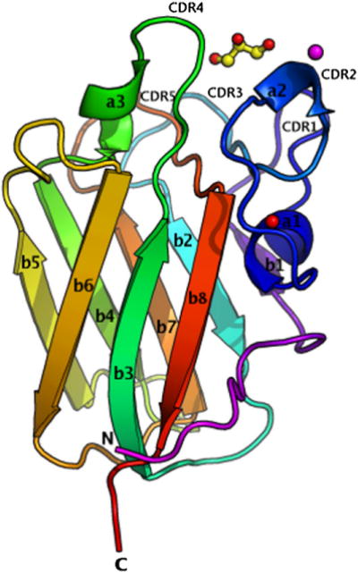
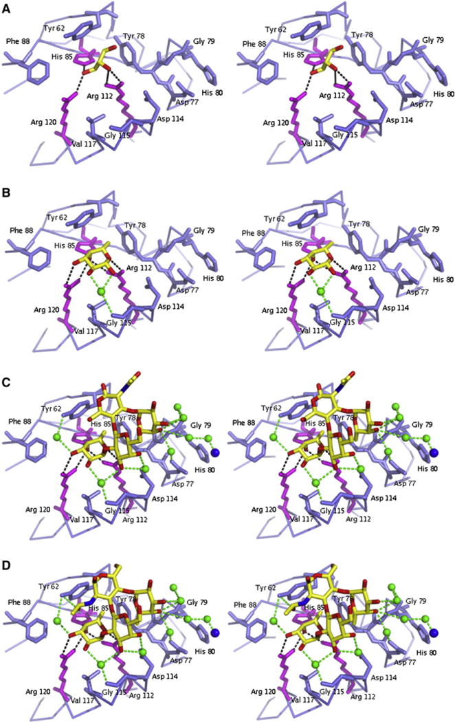
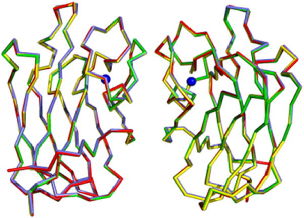
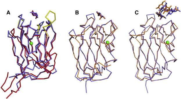
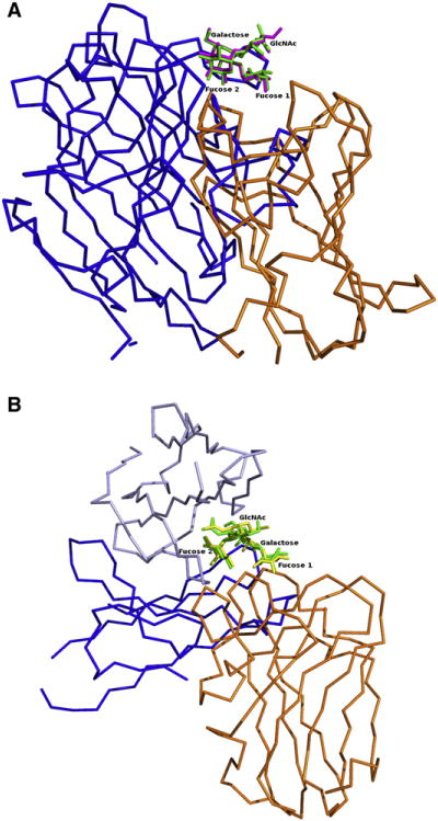
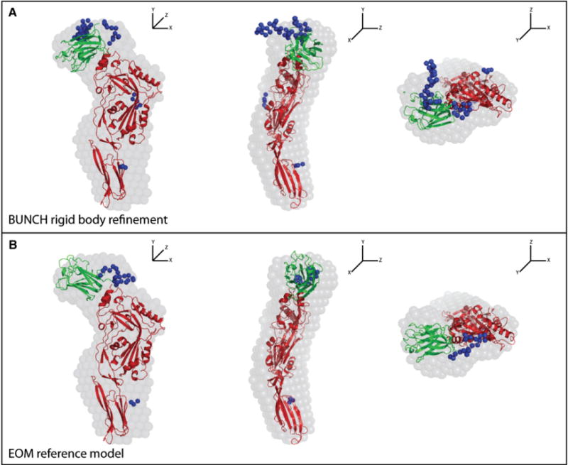
The cholesterol-dependent cytolysins (CDCs) punch holes in target cell membranes through a highly regulated process. Streptococcus mitis lectinolysin (LLY) exhibits another layer of regulation with a lectin domain that enhances the pore-forming activity of the toxin. We have determined the crystal structures of the lectin domain by itself and in complex with various glycans that reveal the molecular basis for the Lewis antigen specificity of LLY. A small-angle X-ray scattering study of intact LLY reveals the molecule is flat and elongated with the lectin domain oriented so that the Lewis antigen-binding site is exposed. We suggest that the lectin domain enhances the pore-forming activity of LLY by concentrating toxin molecules at fucose-rich sites on membranes, thus promoting the formation of prepore oligomers on the surface of susceptible cells.


Figures

Figure 1
Figure 1. Structure of LLYlec
(See also Figure S1, which shows how LLYlec is oriented with respect to the rest of the toxin molecule.) The structure is represented in colors, from the N terminus in magenta to the C terminus in red. Glycerol is shown as ball and stick, the calcium ion is shown as a red sphere, and the Ni ion is shown as a magenta sphere. Secondary structural elements and CDR loops are labeled as described in the text. This and all following structural figures were drawn with PyMol (Delano, 2002).
Figure 2
Figure 2. Close-up Stereo Views of the Ligand Binding Pocket of LLYlec
LLYlec is complexed to (A) glycerol, (B) α-L-fucose, (C) Ley antigen, and (D) Leb antigen. Residues that create the binding pocket are shown as blue and magenta sticks and the ligands as yellow sticks. Water molecules are shown as green spheres, and hydrogen bonds by dashed lines.
Figure 3
Figure 3. Superposition of the Dimers of LLYlec
The dimers are created by crystallographic symmetry. The dimers of LLYlec, LLYlec-fucose, LLYlec-Ley, and LLYlec-Leb are shown in red, yellow, blue, and green, respectively. The Ca2+ ion is shown in blue.
Figure 4
Figure 4. Structural Similarities of LLYlecto Other Proteins
(A) Superposition of the LLYlec domain (in blue) on to the structure of the fucolection of AAA (in red). Both structures are complexed with fucose. The Ca2+ atom, which is located in the same positionin both structures, is shown in green. The CDR loops that are different in AAA, compared to the LLYLec domain, are shown in yellow. (B) Superposition of the fucose complex structures of LLYlec domain (in blue) and the fucolectin module of SpX-1 (in orange). (C) Superposition of the Ley antigen complex structures of LLYlec domain (in blue) and the fucolectin module of SpX-1 (orange).
Figure 5
Figure 5. Protein Recognition of Lewis Antigens
All superpositions are based on the Lewis antigen, and LLYlec is shown in orange. (A) Griffonia simplifolia legume lectin (PDB code: 1LED) is shown in blue with Lewis b antigens superimposed. (B) Monoclonal antibody hu3S193 (PDB code: 1SK3) heavy and light chains are shown in different shades of blue, with Lewis y antigens superimposed.
Figure 6
Figure 6. Comparison of the Ab Initio Shape Envelope with Computationally Docked and SAXS-Derived LLY Models
Each panel shows orthogonal views of the fits. The blue spheres represent missing C-terminal residues and loops between secondary structural elements, including the linker peptide between the lectin domain and the rest of the toxin molecule, which were missing in the ALO structure that was used in the modeling. See also Figure S2 for the SAXS data. (A) Rigid-body refinement of the position of the LLYlec domain with BUNCH (Petoukhov et al., 2005) generated an ensemble of models that were in good agreement with the experimental scattering profile. In all these models, the position of the LLYlec domain corresponded well to the unoccupied density proximal to the N terminus of the LLYCDC domains in the shape envelope. The BUNCH model closest to the average position is shown. (B) As an alternate method of identifying likely LLYlec domain positions, a large pool of models with randomized LLYlec and LLYCDC orientations, constrained by the intervening linker sequence, were generated and interrogated with the EOM (Bernadó et al., 2007). The individual EOM models in the pool with theoretical scattering profiles that best matched the experimental curve were identified. The EOM model closest to the average position is shown.

Comment in

References

    1. Bergelt S, Frost S, Lilie H. Listeriolysin O as cytotoxic component of an immunotoxin. Protein Sci. 2009;18:1210–1220. - PMC - PubMed
    1. Bernadó P, Mylonas E, Petoukhov MV, Blackledge M, Svergun DI. Structural characterization of flexible proteins using small-angle X-ray scattering. J Am Chem Soc. 2007;129:5656–5664. - PubMed
    1. Bianchet MA, Odom EW, Vasta GR, Amzel LM. A novel fucose recognition fold involved in innate immunity. Nat Struct Biol. 2002;9:628–634. - PubMed
    1. Boraston AB, Wang D, Burke RD. Blood group antigen recognition by a Streptococcus pneumoniae virulence factor. J Biol Chem. 2006;281:35263–35271. - PubMed
    1. Boraston AB, Bolam DN, Gilbert HJ, Davies GJ. Carbohydrate-binding modules: fine-tuning polysaccharide recognition. Biochem J. 2004;382:769–781. - PMC - PubMed

Publication types

LinkOut - more resources