

Metabolic engineering of Chinese hamster ovary cells: towards a bioengineered heparin
- PMID: 22326251
- PMCID: PMC3299301
- DOI: 10.1016/j.ymben.2012.01.008
Metabolic engineering of Chinese hamster ovary cells: towards a bioengineered heparin
Abstract
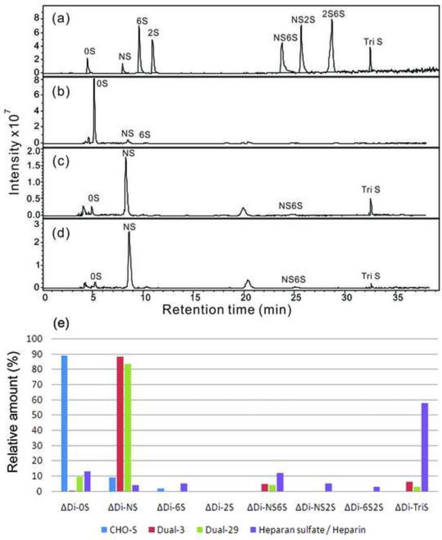
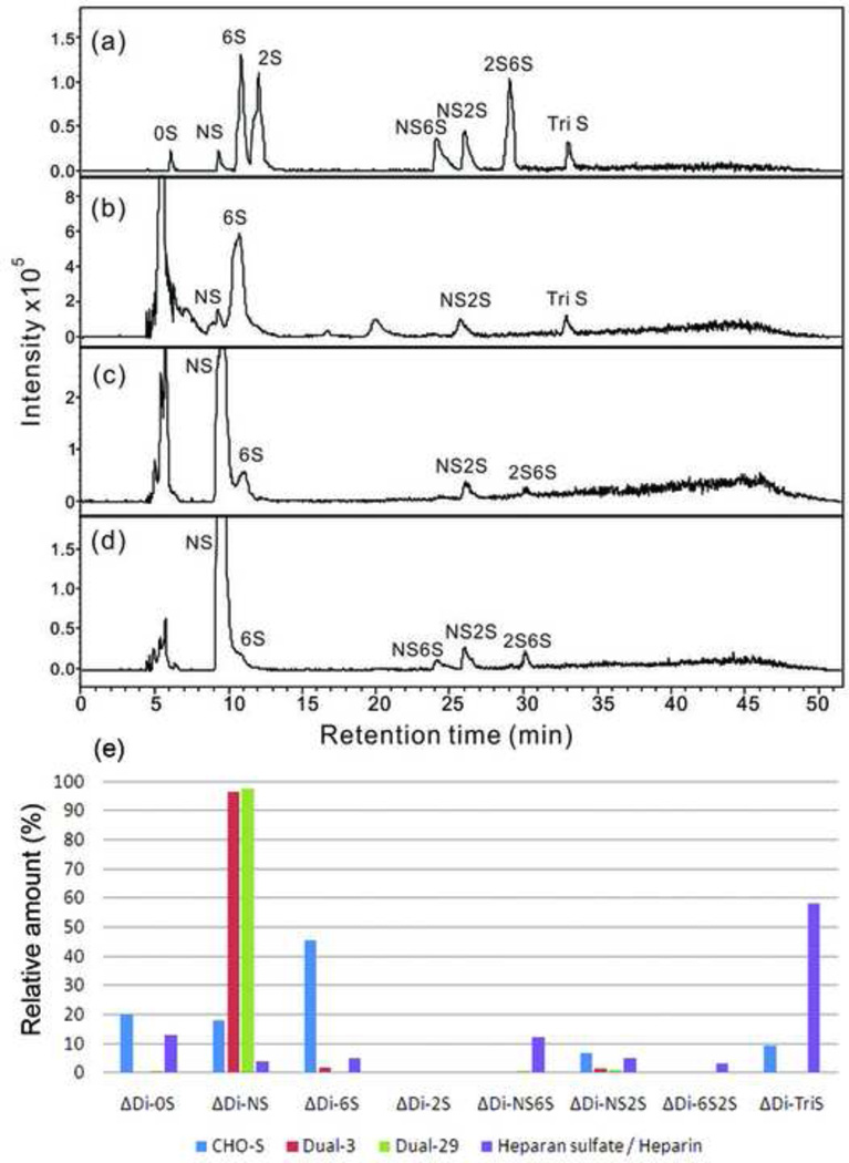
Heparin is the most widely used pharmaceutical to control blood coagulation in modern medicine. A health crisis that took place in 2008 led to a demand for production of heparin from non-animal sources. Chinese hamster ovary (CHO) cells, commonly used mammalian host cells for production of foreign pharmaceutical proteins in the biopharmaceutical industry, are capable of producing heparan sulfate (HS), a related polysaccharide naturally. Since heparin and HS share the same biosynthetic pathway, we hypothesized that heparin could be produced in CHO cells by metabolic engineering. Based on the expression of endogenous enzymes in the HS/heparin pathways of CHO-S cells, human N-deacetylase/N-sulfotransferase (NDST2) and mouse heparan sulfate 3-O-sulfotransferase 1 (Hs3st1) genes were transfected sequentially into CHO host cells growing in suspension culture. Transfectants were screened using quantitative RT-PCR and Western blotting. Out of 120 clones expressing NDST2 and Hs3st1, 2 clones, Dual-3 and Dual-29, were selected for further analysis. An antithrombin III (ATIII) binding assay using flow cytometry, designed to recognize a key sugar structure characteristic of heparin, indicated that Hs3st1 transfection was capable of increasing ATIII binding. An anti-factor Xa assay, which affords a measure of anticoagulant activity, showed a significant increase in activity in the dual-expressing cell lines. Disaccharide analysis of the engineered HS showed a substantial increase in N-sulfo groups, but did not show a pattern consistent with pharmacological heparin, suggesting that further balancing the expression of transgenes with the expression levels of endogenous enzymes involved in HS/heparin biosynthesis might be necessary.
Copyright © 2012 Elsevier Inc. All rights reserved.
Figures


Comment in
-
Toward a bioengineered heparin: challenges and strategies for metabolic engineering of mammalian cells.Bioengineered. 2012 Jul-Aug;3(4):227-31. doi: 10.4161/bioe.20902. Epub 2012 Jun 20. Bioengineered. 2012. PMID: 22714556 Free PMC article.
References
-
- Ahn WS, Antoniewicz MR. Metabolic flux analysis of CHO cells at growth and non-growth phases using isotopic tracers and mass spectrometry. Metab Eng. 2011;13:598–609. - PubMed
-
- Ahsan A, Jeske W, Hoppensteadt D, Lormeau JC, Wolf H, Fareed J. Molecular profiling and weight determination of heparins and depolymerized heparins. J Pharm Sci. 1995;84:724–727. - PubMed
-
- Aldor IS, Keasling JD. Process design for microbial plastic factories: metabolic engineering of polyhydroxyalkanoates. Curr Opin Biotechnol. 2003;14:475–483. - PubMed
-
- Bai X, Esko JD. An animal cell mutant defective in heparan sulfate hexuronic acid 2-O-sulfation. J Biol Chem. 1996;271:17711–17717. - PubMed
Publication types
MeSH terms
Substances
Grants and funding
LinkOut - more resources
Full Text Sources
Other Literature Sources
Medical
