Review
. 2012 Jun;23(4):421-8.
doi: 10.1016/j.semcdb.2012.01.009. Epub 2012 Feb 4.

Notch receptor-ligand binding and activation: insights from molecular studies

Affiliations
Review

Notch receptor-ligand binding and activation: insights from molecular studies

Chandramouli R Chillakuri et al. Semin Cell Dev Biol. 2012 Jun.

Abstract

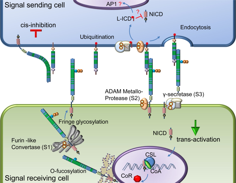
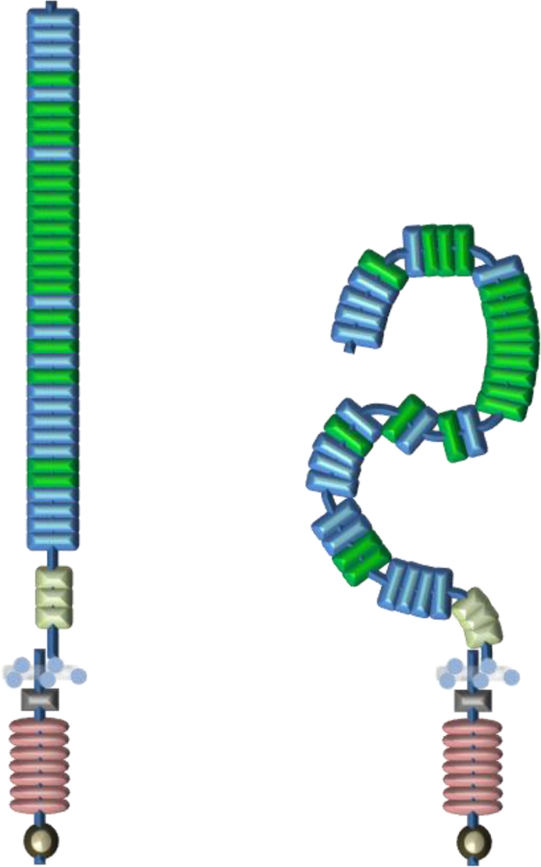
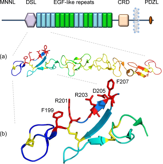
The Notch receptor is part of a core signalling pathway which is highly conserved in all metazoan species. It is required for various cell fate decisions at multiple stages of development and in the adult organism, with dysregulation of the pathway associated with genetic and acquired diseases including cancer. Although cellular and in vivo studies have provided considerable insight into the downstream consequences of Notch signalling, relatively little is known about the molecular basis of the receptor/ligand interaction and initial stages of activation. Recent advances in structure determination of the extracellular regions of human Notch-1 and one of its ligands Jagged-1 have given new insights into docking events occurring at the cell surface which may facilitate the development of new highly specific therapies. We review the structural data available for receptor and ligands and identify the challenges ahead.


Figures

Fig. 1
Fig. 1
Canonical Notch signalling. The Notch receptor undergoes post-translational modifications, including glycosylation and proteolysis (S1) before being targeted to the cell membrane. A ligand from the neighbouring cell binds to the receptor leading to activation. Ubiquitination of ligand and initiation of endocytosis of ligand–receptor complex leads to a second proteolytic cleavage by ADAM metalloprotease (S2) removing the extracellular region. The membrane tethered receptor fragment is cleaved by the γ-secretase complex (S3) to release the Notch intracellular domain (NICD) which translocates into the nucleus. NICD, together with CSL and co-activator Master mind-like (MAML), displaces the co-repressor and forms a transcription activation complex on promoters of target genes like Hes-1, Hes-5 which contain CSL binding sites. Some recent reports show that ligands also undergo proteolysis and release ligand intracellular domain (LICD) which antagonizes Notch signalling by mechanisms as yet unclear. Receptor and ligand present on the same cell surface can also bind to each other leading to cis-inhibition. The molecular basis of the cis-inhibitory complex is unknown, but implicates similar regions of receptor and ligand to those involved in trans-activation.
Fig. 2
Fig. 2
Architecture of Notch1 receptor. Human Notch-1 is represented with major domains annotated. In the EGF repeat region the Ca2+ binding EGF domain is green and non-Ca2+ binding EGF domain is blue. (a) Crystal structure of EGF11–13 repeats (PDB ID: 2VJ2) which encompass the ligand binding site and show a near linear domain arrangement. (b) Tandem EGF repeats are rigidified by bound Ca2+ (red sphere) and interdomain packing of aromatic residues, Tyrosine (purple spheres) from EGF11 with hydrophobic Isoleucine (blue spheres) and Glycine (green spheres) from EGF12. (c) Crystal structure of the negative regulatory region (NRR) (PDB ID: 3I08) comprising three LNR repeats and the heterodimerisation domain shows that the S2 cleavage site is buried deep inside the globular NRR. (d) Crystal structure of ankyrin repeat region ANK (PDB ID: 2F8Y). (e) Transcription activation complex (PDB ID: 2F8X) made up of CSL (green), NICD (blue) and Master mind-like (purple) binding to the CSL binding site (double stranded DNA) on Hes-1 promoter. RAM peptide region involved in CSL protein binding, trans-activating domain (TAD) and degradation signal PEST are also illustrated in the figure.
Fig. 3
Fig. 3
Cell surface organisation of Notch receptor. Much of the extracellular region of the Notch receptor is made up of EGF repeats. The crystal structure of the EGF11–13 region showed that these domains are arranged into a near linear structure rigidified by Ca2+. Based on this observation, a near linear model is proposed that extends away from the membrane. There is evidence that the cbEGF–EGF and EGF–EGF tandem domains can be flexible or pack in a non-linear orientation and may adopt a bent rather than a linear structure. Since the N-terminal region and C-terminal region of the extracellular domain (ECD) contains several non Ca2+ binding EGF repeats, it is possible that the Notch receptor attains a much more compact structure. It should be noted that the compact structure represented here is showing possible regions of flexibility and is not a structure prediction. If there are any interactions between different regions of the ECD a much more compact structure may exist.
Fig. 4
Fig. 4
Architecture of Jagged1. Human Jagged-1 is represented in the figure. Similar to the Notch receptor, much of the extracellular region comprises EGF repeats. No structural predictions exist for the N-terminus of Notch ligand (MNNL) region. CRD represents the cysteine-rich region and the PDZL domain is present on the C-terminal region. (a) Crystal structure of human Jagged1 DSL EGF3 region (PDB ID: 2VJ3) showed a near linear arrangement of these domains. Disulphide bonds are shown as yellow sticks (b) The DSL domain is found to have a unique fold and forms the main receptor binding site. Sequence alignments and subsequent mutational studies showed that a subset of residues localised on one face of Jagged1 are crucial for the interactions with the receptor. Side chains of residues F199, R201, R203, D205, F207 are represented as red sticks.

References

    1. Kopan R., Ilagan M.X. The canonical Notch signalling pathway: unfolding the activation mechanism. Cell. 2009;137:216–233. - PMC - PubMed
    1. Sanchez-Irizarry C., Carpenter A.C., Weng A.P., Pear W.S., Aster J.C., Blacklow S.C. Notch subunit heterodimerization and prevention of ligand-independent proteolytic activation depend, respectively on a novel domain and the LNR repeats. Mol Cell Biol. 2004;24:9265–9273. - PMC - PubMed
    1. Tamura K., Taniguchi Y., Minoguchi S., Sakai T., Tun T., Furukawa T. Physical interaction between a novel domain of the receptor Notch and the transcription factor RBO-J kappa/Su(H) Curr Biol. 1995;5:1416–1423. - PubMed
    1. Weng A.P., Ferrando A.A., Lee W., Morris J.P., 4th, Silverman L.B., Sanchez-Irizarry C. Activating mutations of Notch1 in human T cell acute lymphoblastic leukemia. Science. 2004;306:269–271. - PubMed
    1. Rebay I., Flemming R.J., Fehon R.G., Cherbas L., Cherbas P., Artavanis-Tsakonas S. Specific EGF repeats of Notch mediate interactions with Delta and Serrate: implications for Notch as a multifunctional receptor. Cell. 1991;67:687–699. - PubMed

Publication types

MeSH terms

LinkOut - more resources