

Gene deletions and amplifications in human hepatocellular carcinomas: correlation with hepatocyte growth regulation
- PMID: 22326833
- PMCID: PMC3657620
- DOI: 10.1016/j.ajpath.2011.12.021
Gene deletions and amplifications in human hepatocellular carcinomas: correlation with hepatocyte growth regulation
Abstract
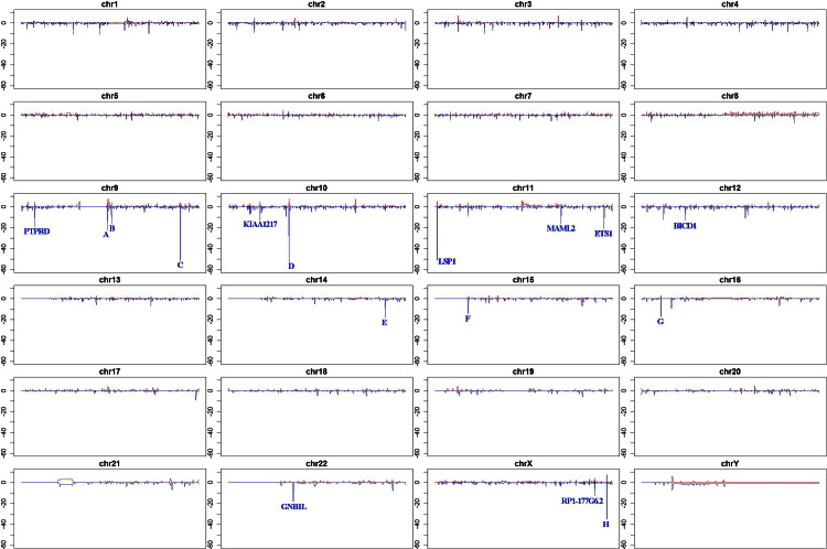
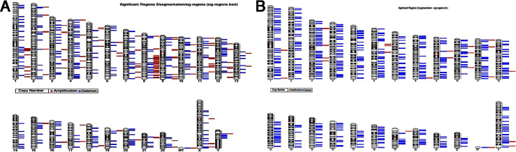
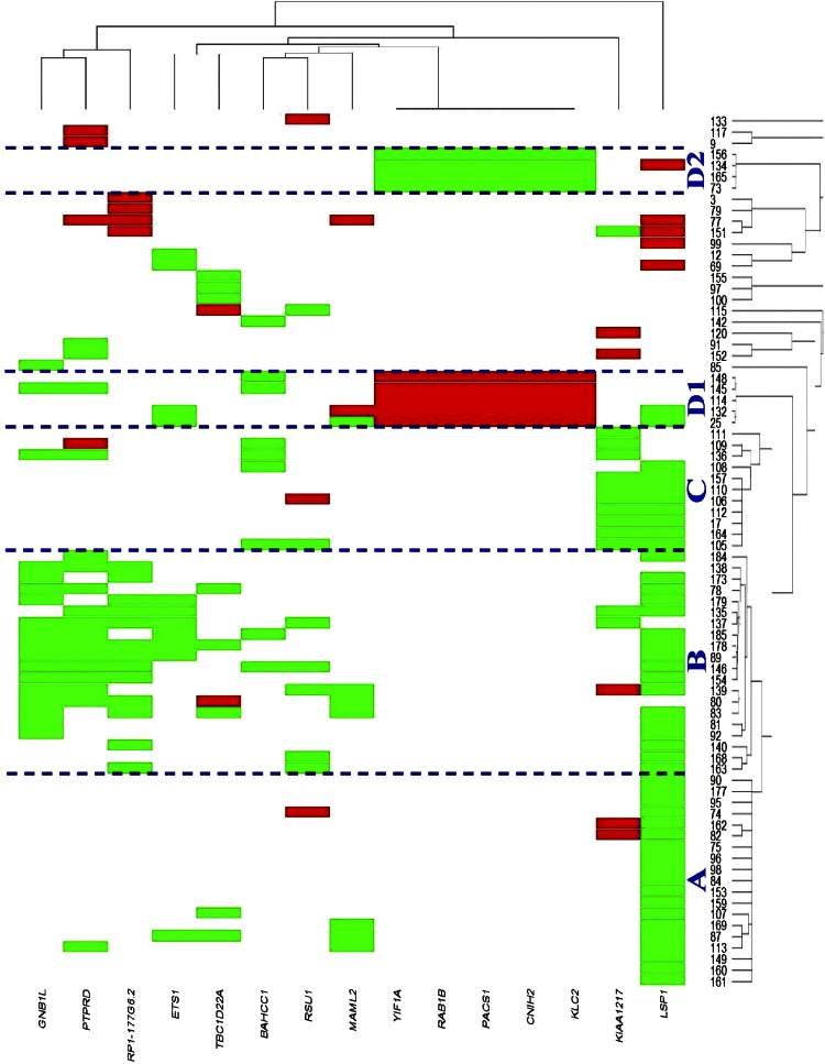
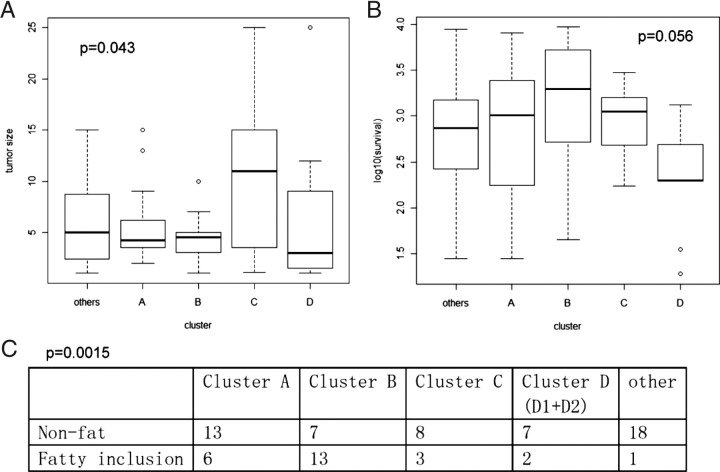
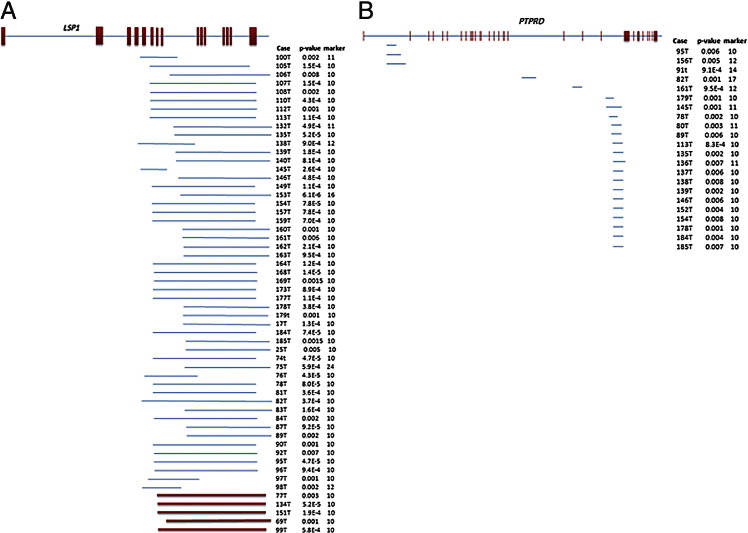
Tissues from 98 human hepatocellular carcinomas (HCCs) obtained from hepatic resections were subjected to somatic copy number variation (CNV) analysis. Most of these HCCs were discovered in livers resected for orthotopic transplantation, although in a few cases, the tumors themselves were the reason for the hepatectomies. Genomic analysis revealed deletions and amplifications in several genes, and clustering analysis based on CNV revealed five clusters. The LSP1 gene had the most cases with CNV (46 deletions and 5 amplifications). High frequencies of CNV were also seen in PTPRD (21/98), GNB1L (18/98), KIAA1217 (18/98), RP1-1777G6.2 (17/98), ETS1 (11/98), RSU1 (10/98), TBC1D22A (10/98), BAHCC1 (9/98), MAML2 (9/98), RAB1B (9/98), and YIF1A (9/98). The existing literature regarding hepatocytes or other cell types has connected many of these genes to regulation of cytoskeletal architecture, signaling cascades related to growth regulation, and transcription factors directly interacting with nuclear signaling complexes. Correlations with existing literature indicate that genomic lesions associated with HCC at the level of resolution of CNV occur on many genes associated directly or indirectly with signaling pathways operating in liver regeneration and hepatocyte growth regulation.
Copyright © 2012 American Society for Investigative Pathology. Published by Elsevier Inc. All rights reserved.
Figures
References
-
- Pitot H.C. Adventures in hepatocarcinogenesis. Ann Rev Pathol. 2007;2:1–29. - PubMed
-
- Thomas M.B., Jaffe D., Choti M.M., Belghiti J., Curley S., Fong Y., Gores G., Kerlan R., Merle P., O'Neil B., Poon R., Schwartz L., Tepper J., Yao F., Haller D., Mooney M., Venook A. Hepatocellular carcinoma: consensus recommendations of the National Cancer Institute Clinical Trials Planning Meeting. J Clin Oncol. 2010;28:3994–4005. - PMC - PubMed
-
- Feitelson M.A., Reis H.M., Liu J., Lian Z., Pan J. Hepatitis B virus X antigen (HBxAg) and cell cycle control in chronic infection and hepatocarcinogenesis. Front Biosci. 2005;10:1558–1572. - PubMed
-
- Lee J.S., Thorgeirsson S.S. Genetic profiling of human hepatocellular carcinoma. Sem Liver Dis. 2005;25:125–132. - PubMed
-
- Lee J.S., Chu I.S., Heo J., Calvisi D.F., Sun Z., Roskams T., Durnez A., Demetris A.J., Thorgeirsson S.S. Classification and prediction of survival in hepatocellular carcinoma by gene expression profiling. Hepatology. 2004;40:667–676. - PubMed
Publication types
MeSH terms
Substances
Grants and funding
LinkOut - more resources
Full Text Sources
Medical
Miscellaneous
