. 2012 Mar;122(3):1109-18.
doi: 10.1172/JCI60329. Epub 2012 Feb 13.

Metabolic stress-induced activation of FoxO1 triggers diabetic cardiomyopathy in mice

Affiliations

Metabolic stress-induced activation of FoxO1 triggers diabetic cardiomyopathy in mice

Pavan K Battiprolu et al. J Clin Invest. 2012 Mar.

Abstract

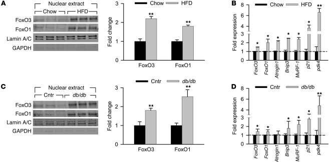
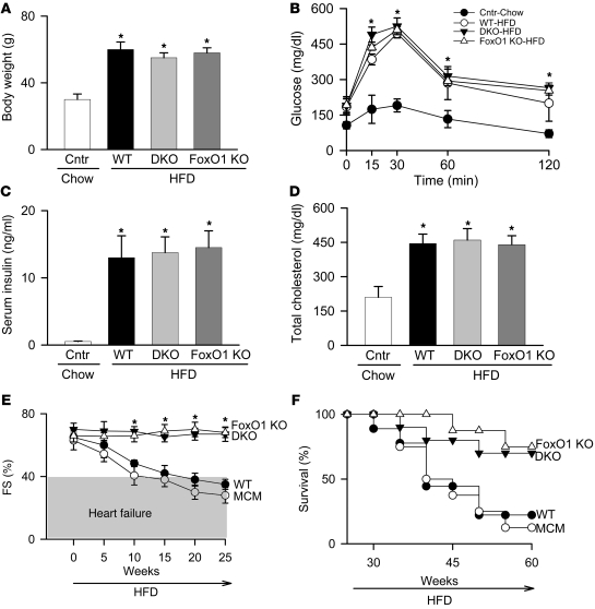
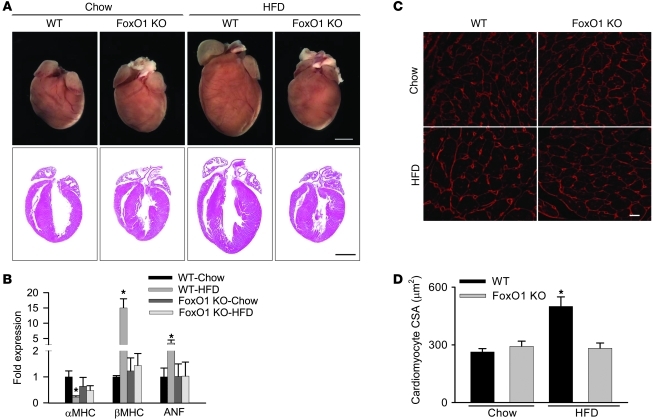
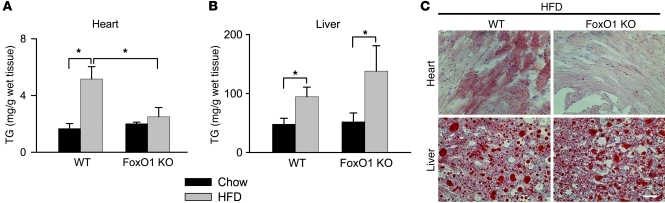
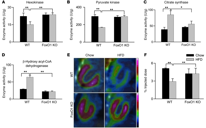
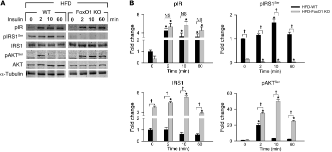
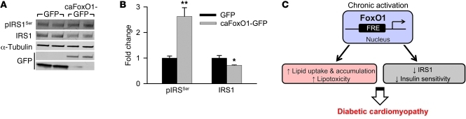
The leading cause of death in diabetic patients is cardiovascular disease; diabetic cardiomyopathy is typified by alterations in cardiac morphology and function, independent of hypertension or coronary disease. However, the molecular mechanism that links diabetes to cardiomyopathy is incompletely understood. Insulin resistance is a hallmark feature of diabetes, and the FoxO family of transcription factors, which regulate cell size, viability, and metabolism, are established targets of insulin and growth factor signaling. Here, we set out to evaluate a possible role of FoxO proteins in diabetic cardiomyopathy. We found that FoxO proteins were persistently activated in cardiac tissue in mice with diabetes induced either genetically or by high-fat diet (HFD). FoxO activity was critically linked with development of cardiomyopathy: cardiomyocyte-specific deletion of FoxO1 rescued HFD-induced declines in cardiac function and preserved cardiomyocyte insulin responsiveness. FoxO1-depleted cells displayed a shift in their metabolic substrate usage, from free fatty acids to glucose, associated with decreased accumulation of lipids in the heart. Furthermore, we found that FoxO1-dependent downregulation of IRS1 resulted in blunted Akt signaling and insulin resistance. Together, these data suggest that activation of FoxO1 is an important mediator of diabetic cardiomyopathy and is a promising therapeutic target for the disease.


Figures

Figure 1
Figure 1. FoxOs are persistently active in HFD and db/db hearts.
(A and B) For HFD studies, hearts were collected from mice fed HFD starting at 8 weeks of age for 10 weeks and compared with age-matched chow diet–fed mice. (C and D) For db/db studies, hearts were collected from 13- to 15-week-old db/db mice and compared with age-matched controls (Cntr; db/+ and WT combined). (A and C) Immunoblot detection of FoxO3 and FoxO1 proteins from the nuclear extract. Quantification of band density normalized to lamin A/C is also shown. (B and D) mRNA expression of FoxO downstream target genes using real-time RT-PCR, normalized to 18S RNA. n = 5 per group. *P < 0.05; **P < 0.01 versus respective control group.
Figure 2
Figure 2. FoxOs are required for HFD-induced cardiac dysfunction and mortality.
Body weight (A), oral glucose tolerance (B), serum insulin (C), total cholesterol (D), percent FS (E), and survival (F) in WT, DKO, and FoxO1 KO mice fed HFD for 25 weeks (AE) or longer (F) and in normal chow–fed controls (Cntr; respective genotypes combined). (B) Mice were fasted for 16 hours prior to the oral glucose challenge. (E) Heart failure was defined as FS ≤ 40%. (F) Survival curve was generated using at least 12 mice per group. *P < 0.05 versus chow-fed control group.
Figure 3
Figure 3. FoxOs are essential for HFD-induced cardiac hypertrophy.
Gross morphology (A) and mRNA expression of fetal gene markers, assessed by real-time RT-PCR and normalized to 18S RNA (B), in hearts from WT and FoxO1 KO mice fed either chow or HFD. (C and D) Hearts from HFD-fed WT mice demonstrated larger cardiomyocyte CSA. (C) Representative images of LV cardiomyocytes (obtained from a transverse section of LV) stained with wheat germ agglutinin (red). (D) Cardiomyocyte CSA was quantified from at least 15 cells per section and 3 sections per group. Scale bars: 2 mm (A); 20 μm (C). n = 5. *P < 0.05 versus chow-fed WT and HFD-fed FoxO1 KO.
Figure 4
Figure 4. FoxOs are required for HFD-induced lipotoxicity.
TG content (A and B) and lipid accumulation, evidenced by Oil Red O staining (C), were elevated in both hearts and livers from WT animals, and only in livers from FoxO1 KO mice fed HFD. Scale bar: 100 μm. n = 3–5. *P < 0.05.
Figure 5
Figure 5. HFD-induced changes in cardiac metabolism require FoxO1.
Hearts from HFD-fed WT mice manifested lower glycolytic activity (A and B), higher oxidative activity (C and D), and reduced glucose uptake (E and F) compared with chow-fed WT and HFD-fed FoxO1 KO mouse hearts. (AD) Maximal activities of the glycolytic enzymes hexokinase (A) and pyruvate kinase (B) and the oxidative enzymes citrate synthase (C) and β-hydroxyl acyl-CoA dehydrogenase (D). (E) Myocardial glucose uptake, visualized by FDG PET. (F) FDG uptake was proportional to the intensity of radioactive contrast, quantified as the radioactivity in a myocardial region-of-interest and expressed as a percentage of the total injected dose. n = 3–5. *P < 0.05, **P < 0.01 as indicated by brackets.
Figure 6
Figure 6. Sustained insulin sensitivity in FoxO1 KO hearts is regulated at the level of IRS1 and Akt phosphorylation.
(A) Immunoblot detection of proteins involved in the insulin signaling cascade. (B) Quantification of band density normalized to α-tubulin. Proteins were studied from whole cell extracts of isolated adult cardiomyocytes treated with insulin (100 nM) for 0, 2, 10, or 60 minutes. pIR, phosphorylated IRβ (Tyr1150/1151 residue); pAKTSer, phosphorylated Akt (Ser473 residue). n = 3. *P < 0.05 versus 0 minutes; P < 0.05 as indicated by brackets.
Figure 7
Figure 7. Constitutive expression of FoxO1 inactivates IRS1.
(A) Immunoblot detection of pIRS1Ser and IRS1 in isolated neonatal ventricular cardiomyocytes overexpressing GFP or GFP-tagged caFoxO1. (B) Quantification of band density normalized to α-tubulin. n = 3. *P < 0.05, **P < 0.01 versus GFP. (C) Working model of the role of FoxO1 in insulin resistance–induced diabetic cardiomyopathy. FRE, FoxO response element.

References

    1. Roger VL, et al. Heart disease and stroke statistics--2011 update: a report from the American Heart Association. Circulation. 2011;123(4):e18–e209. doi: 10.1161/CIR.0b013e3182009701. - DOI - PMC - PubMed
    1. Witteles RM, Fowler MB. Insulin-resistant cardiomyopathy clinical evidence, mechanisms, and treatment options. J Am Coll Cardiol. 2008;51(2):93–102. doi: 10.1016/j.jacc.2007.10.021. - DOI - PubMed
    1. Battiprolu PK, Gillette TG, Wang ZV, Lavandero S, Hill JA. Diabetic cardiomyopathy: mechanisms and therapeutic targets. Drug Discov Today Dis Mech. 2010;7(2):e135–e143. doi: 10.1016/j.ddmec.2010.08.001. - DOI - PMC - PubMed
    1. Ferdous A, Battiprolu PK, Ni YG, Rothermel BA, Hill JA. FoxO, autophagy, and cardiac remodeling. J Cardiovasc Transl Res. 2010;3(4):355–364. doi: 10.1007/s12265-010-9200-z. - DOI - PMC - PubMed
    1. Cheng Z, White MF. Targeting Forkhead box O1 from the concept to metabolic diseases: lessons from mouse models. Antioxid Redox Signal. 2011;14(4):649–661. doi: 10.1089/ars.2010.3370. - DOI - PMC - PubMed

Publication types