. 2012 Apr 4;31(7):1679-91.
doi: 10.1038/emboj.2012.18. Epub 2012 Feb 10.

IAPs limit activation of RIP kinases by TNF receptor 1 during development

Affiliations

IAPs limit activation of RIP kinases by TNF receptor 1 during development

Maryline Moulin et al. EMBO J. .

Abstract

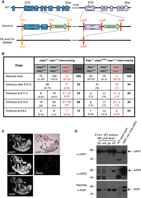
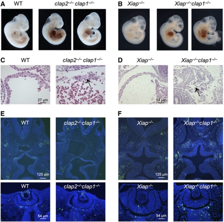
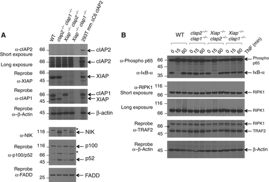
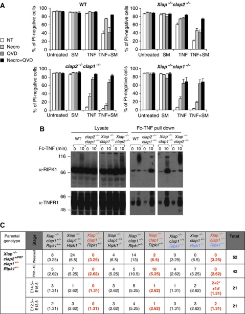
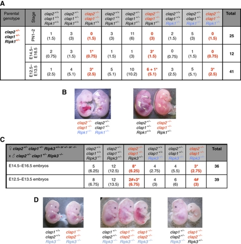
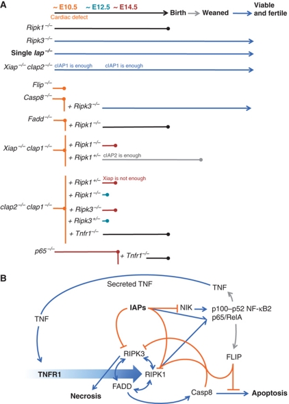
Inhibitor of apoptosis (IAP) proteins cIAP1, cIAP2, and XIAP (X-linked IAP) regulate apoptosis and cytokine receptor signalling, but their overlapping functions make it difficult to distinguish their individual roles. To do so, we deleted the genes for IAPs separately and in combination. While lack of any one of the IAPs produced no overt phenotype in mice, deletion of cIap1 with cIap2 or Xiap resulted in mid-embryonic lethality. In contrast, Xiap(-/-)cIap2(-/-) mice were viable. The death of cIap2(-/-)cIap1(-/-) double mutants was rescued to birth by deletion of tumour necrosis factor (TNF) receptor 1, but not TNFR2 genes. Remarkably, hemizygosity for receptor-interacting protein kinase 1 (Ripk1) allowed Xiap(-/-)cIap1(-/-) double mutants to survive past birth, and prolonged cIap2(-/-)cIap1(-/-) embryonic survival. Similarly, deletion of Ripk3 was able to rescue the mid-gestation defect of cIap2(-/-)cIap1(-/-) embryos, as these embryos survived to E15.5. cIAPs are therefore required during development to limit activity of RIP kinases in the TNF receptor 1 signalling pathway.


Conflict of interest statement

DLV and JS are on the Scientific Advisory Board of TetraLogic Pharmaceuticals.

Figures

Figure 1
Figure 1
Deletion of cIap1 plus cIap2, or cIap1 plus Xiap, results in embryonic lethality at ∼E10. (A) Generation of a genetically modified cIap locus. Exons 2 and 3 of cIap2 were flanked by frt sites and a neomycin phosphotransferase expression cassette and exon 1 of cIap1 was flanked by loxP sites with an hygromycin cassette. (B) Incidence of genotypes of weaned mice or embryos derived from indicated intercrossed mice; * represents embryos without heartbeat or reabsorbed and # represents embryos with minimal heart contractile activity. Expected numbers of each group shown in brackets (based on Mendelian ratios). (C) Detection of cIap1, cIap2 and Xiap expression during development (E10.5) by in situ hybridization as compared with sense control probe. (D) Expression of IAPs during mouse development detected by western blotting. A WT E10.5 embryo was lysed in DISC lysis buffer supplemented with protease inhibitors, NEM and Pefabloc. IAPs were trapped using biotinylated SM and precipitated with streptavidin beads. Lysates (lys), unbound fractions (unb), glycine elutions (ge), and boiled beads (bb) were separated using SDS/PAGE and blotted with antibodies to cIAP1, cIAP2, and XIAP. 293T cells transfected with a plasmid encoding mouse cIAP2ΔC6 were used as a positive control for detection of cIAP2. See also Supplementary Figure S1. Figure source data can be found in Supplementary data.
Figure 2
Figure 2
Embryonic lethality of cIap2−/−cIap1−/− and Xiap−/−cIap1−/− DKO embryos. (A, B) Whole view of E10.5 and E11.5 embryos derived from intercrosses of cIap2+/−cIap1+/− and Xiap−/−cIap1+/− mice, respectively. In the DKO embryos, blood has accumulated in the pericardial cavities, but not in those of WT or Xiap−/− littermates. (C, D) Histological analysis of the atria from a WT and cIap2−/−cIap1−/− DKO E10.5 embryo and Xiap−/− and Xiap−/−cIap1−/− DKO E11.5 embryos. The arrow shows discontinuity in the wall. (E, F) TUNEL (green) staining indicating fragmented DNA and nuclear (DAPI, blue) staining of E10.5–E11.5 transverse sections of embryos with genotypes as indicated. Heads are shown in the top panels and eyes are shown in the bottom panels. No gross differences in patterns of TUNEL-stained cells were observed.
Figure 3
Figure 3
Defects in NF-κB signalling in the absence of cIAP1. (A) Levels of NIK, and spontaneous processing of p100 are increased in MEFs lacking cIAP1. SV40 large T antigen-immortalized MEFs derived from WT, cIap2−/−cIap1−/− DKO, Xiap−/−cIap2−/− DKO, and Xiap−/−cIap1−/− DKO embryos, were lysed then separated using SDS/PAGE and probed with antibodies to cIAP1, cIAP2, XIAP, NIK, p100/p52, and FADD (* indicates non-specific bands due to the antibody to NIK). Lysate from 293T cells transfected with a plasmid encoding murine cIAP2 ΔC6 was used as a positive control for detection of cIAP2. (B) TNF does not efficiently trigger IκB degradation in cIap1 deleted cells. Cells described in (A) were incubated with or without 100 ng/ml Fc-TNF for the indicated times. Lysates were separated using SDS/PAGE and probed for phosphorylated p65, IκBα, RIPK1, and TRAF2 and β-actin. Figure source data can be found in Supplementary data.
Figure 4
Figure 4
Endogenous levels of cIAP1, and ectopic expression of cIAP2, protect MEFs from killing by TNF much more than XIAP. (A) Sensitivity of gene deleted lines to induction of cell death. Cells described in Figure 3A were incubated for 24 h with 100 ng/ml Fc-TNF or 20 ng Fc-CD95L, 500 nM SM, 1 μM etoposide or 5 μg/ml cisplatin where indicated. Cells were stained with PI and analysed by flow cytometry. The mean+s.e.m. of 3–11 independent experiments is shown. (B, C) Ability of ectopically expressed IAPs to protect cIap2−/−cIap1−/− DKO cells. cIap2−/−cIap1−/− MEFs were complemented with inducible mouse cIAP1, FLAG–cIAP2, or mouse XIAP and induced with 10 nM 4-HT for 24 h, and then incubated with or without 100 ng/ml Fc-TNF for a further 24 h. Cells were stained with PI and analysed by flow cytometry. The mean values+s.e.m. of 5–14 independent experiments are shown (* indicates a non-specific band). (D, E) Ability of ectopically expressed IAPs to protect Xiap−/−cIap1−/− DKO cells. Xiap−/−cIap1−/− MEFs were complemented with inducible mouse cIAP1 or EGFP–XIAP, induced with 10 nM 4-HT for 24 h and then treated with 100 ng/ml Fc-TNF or 20 ng/ml Fc-CD95L and 500 nM SM. The mean values+s.e.m. of three to six independent experiments are shown. See also Supplementary Figure S2. Figure source data can be found in Supplementary data.
Figure 5
Figure 5
Deletion of Tnfr1, but not Tnfr2 allows cIap2−/−cIap1−/− embryos to develop until birth. (A) Incidence of genotypes of weaned mice or embryos derived from intercrosses of genotypes as indicated; * represents embryos without heartbeat or reabsorbed. Expected numbers of each group are shown in brackets. (B) Representative photographs of three 1-day-old pups from a litter from a cIap2+/−cIap1+/−Tnfr1−/− intercross. (C) Sensitivity to TNF is conferred by TNFR1 not TNFR2. SV40 large T antigen transformed MEFs derived from WT embryos or mutant embryos as indicated were incubated for 24 h in the presence or absence of 100 ng/ml Fc-TNF and 500 nM SM. Cells were stained with PI and analysed by flow cytometry. The mean values+s.e.m. of three to seven independent experiments are shown.
Figure 6
Figure 6
In the absence of IAPs, both TNF-induced death of MEFs, and embryonic death during development, involves RIPK1. (A) Together, the RIPK1 kinase inhibitor necrostatin and the caspase inhibitor QVD protect IAP gene deleted MEFs from killing by TNF. MEFs derived from WT and compound mutant embryos were incubated for 24 h with or without 100 ng/ml Fc-TNF and 500 nM SM in the presence or absence of 50 μM necrostatin or 10 μM QVD. Cells were stained with PI and analysed by flow cytometry. The mean values+s.e.m. of 3–11 independent experiments are shown. (B) TNF-induced RIPK1 modification fails, and receptor-associated RIPK1 is increased when cIap1 genes are deleted. MEFs were stimulated with 1 μg/ml Fc-TNF prior to Fc pull down, and analysed by western blotting. (C) Incidence genotypes of offspring from Xiap−/−cIap1+/−Ripk1+/− intercrosses. *Represents embryos without heartbeat or reabsorbed and # represents embryos alive but with a small liver. See also Supplementary Figure S3. Figure source data can be found in Supplementary data.
Figure 7
Figure 7
Prevention of E10.5 lethality of cIap2−/−cIap1−/− embryos by deletion of Ripk1 or Ripk3 genes. (A) Incidence of genotypes of born pups or embryos derived from intercrosses of cIap2+/−cIap1+/−Ripk1+/− mice. (B) Representative photographs of E12.5 embryos from a cIap2+/−cIap1+/−Ripk1+/− intercross. (C) Incidence of genotypes of embryos derived from crosses of cIap2+/−cIap1+/−Ripk3+/+or+/−or−/− females with cIap2+/−cIap1+/−Ripk3−/− males. In (A, C), * represents embryos without heartbeat or reabsorbed and # represents embryos alive but with a small liver. (D) Representative photographs of E13.5 and E14.5 embryos from a cIap2+/−cIap1+/−Ripk3+/− female with cIap2+/−cIap1+/−Ripk3−/− male cross.
Figure 8
Figure 8
Roles of cIAPs in signalling and cell death during development. (A) Diagram of extent of viability of single, double, and triple gene deleted mice. (B) Speculative model to account for the phenotypes of gene deleted mice. Binding of TNF to TNFR1 triggers (large blue arrow) formation of complexes that can culminate in cell death by apoptosis or necroptosis, or lead to cell survival. Activating interactions are indicated with blue arrows; inhibitory interactions are indicated with orange lines; transcriptional induction is indicated with grey arrows. Merging of lines (such as those from Casp8 and FLIP to RIPK1, or IAPs and RIPK1 to p65/RelA) indicate proteins that can act together. cIAPs limit levels of NIK, and inhibit cell death mediated by RIPK1 and RIPK3, but cooperate with RIPK1 to activate p65/RelA. RIPK1 has both pro-death and pro-survival functions, by promoting necroptosis via RIPK3, apoptosis via FADD and Casp8, and cell survival via p65/RelA. Casp8 inhibits cell death by necrosis, but promotes cell death by apoptosis. FLIP inhibits cell death by both pathways. This model is not complete, and does not include other important proteins, such as TRADD, TRAF2, CYLD, A20, TAK1, HOL, HOIP, or Sharpin, just to name a few. For further details, see Discussion.

Comment in

  • cIAP2 supports viability of mice lacking cIAP1 and XIAP.
    Heard KN, Bertrand MJ, Barker PA. Heard KN, et al. EMBO J. 2015 Oct 1;34(19):2393-5. doi: 10.15252/embj.201592060. EMBO J. 2015. PMID: 26427758 Free PMC article. No abstract available.
  • Response to Heard et al.
    Moulin M, Voss AK, Thomas T, Wong WW, Cook WD, Koentgen F, Vince J, Silke J, Vaux DL. Moulin M, et al. EMBO J. 2015 Oct 1;34(19):2396-7. doi: 10.15252/embj.201592761. EMBO J. 2015. PMID: 26427759 Free PMC article. No abstract available.

References

    1. Bertrand MJ, Milutinovic S, Dickson KM, Ho WC, Boudreault A, Durkin J, Gillard JW, Jaquith JB, Morris SJ, Barker PA (2008) cIAP1 and cIAP2 facilitate cancer cell survival by functioning as E3 ligases that promote RIP1 ubiquitination. Mol Cell 30: 689–700 - PubMed
    1. Birnbaum MJ, Clem RJ, Miller LK (1994) An apoptosis inhibiting gene from a nuclear polyhedrosis virus encoding a polypeptide with cys/his sequence motif. J Virol 68: 2521–2528 - PMC - PubMed
    1. Bossen C, Ingold K, Tardivel A, Bodmer JL, Gaide O, Hertig S, Ambrose C, Tschopp J, Schneider P (2006) Interactions of tumor necrosis factor (TNF) and TNF receptor family members in the mouse and human. J Biol Chem 281: 13964–13971 - PubMed
    1. Caserta TM, Smith AN, Gultice AD, Reedy MA, Brown TL (2003) Q-VD-OPh, a broad spectrum caspase inhibitor with potent antiapoptotic properties. Apoptosis 8: 345–352 - PubMed
    1. Ch’en IL, Tsau JS, Molkentin JD, Komatsu M, Hedrick SM (2011) Mechanisms of necroptosis in T cells. J Exp Med 208: 633–641 - PMC - PubMed

Publication types

MeSH terms

Substances