Review
. 2012 Mar;123(3):295-319.
doi: 10.1007/s00401-012-0954-z. Epub 2012 Feb 12.

Pathology of peripheral nerve sheath tumors: diagnostic overview and update on selected diagnostic problems

Affiliations
Review

Pathology of peripheral nerve sheath tumors: diagnostic overview and update on selected diagnostic problems

Fausto J Rodriguez et al. Acta Neuropathol. 2012 Mar.

Abstract

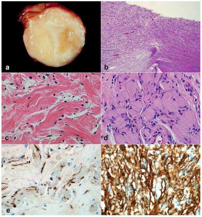
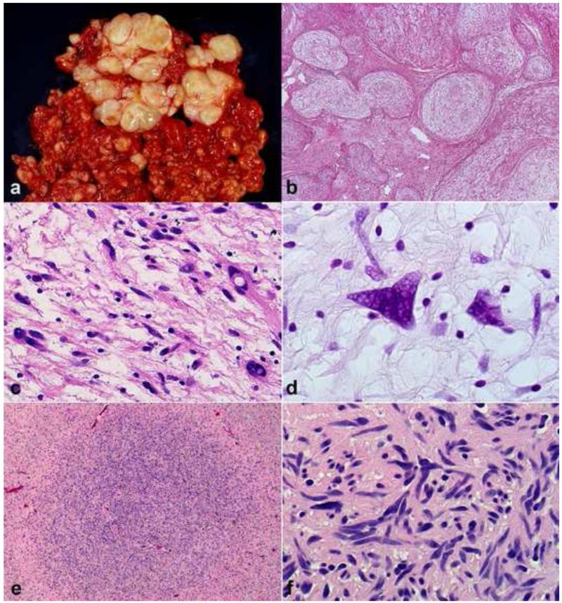
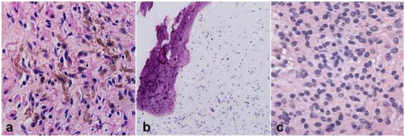
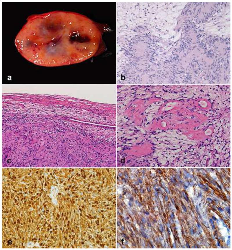
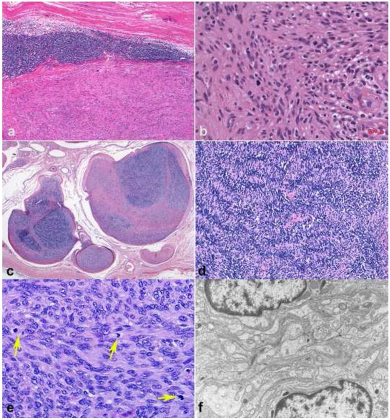
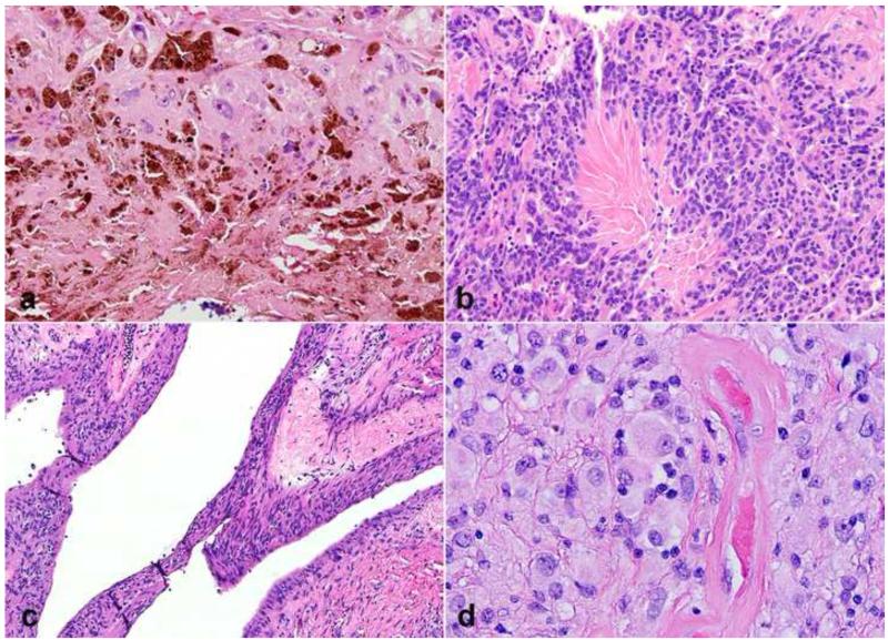
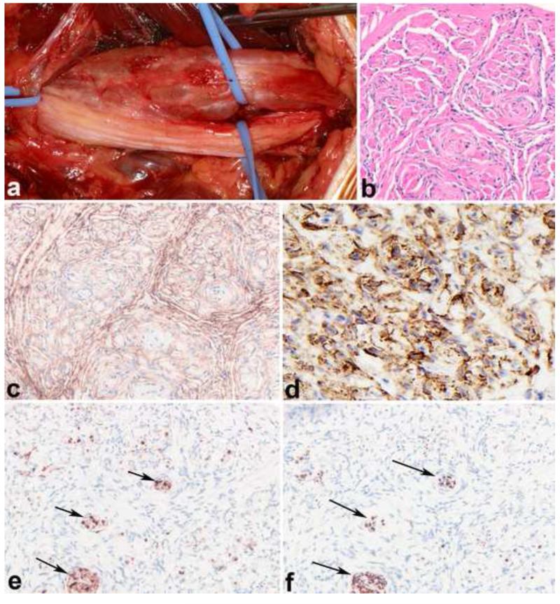
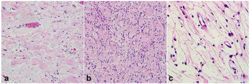
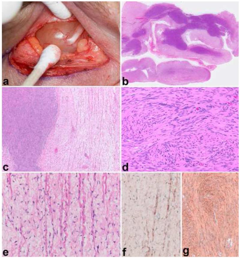
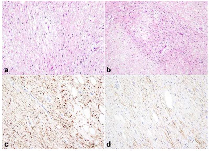
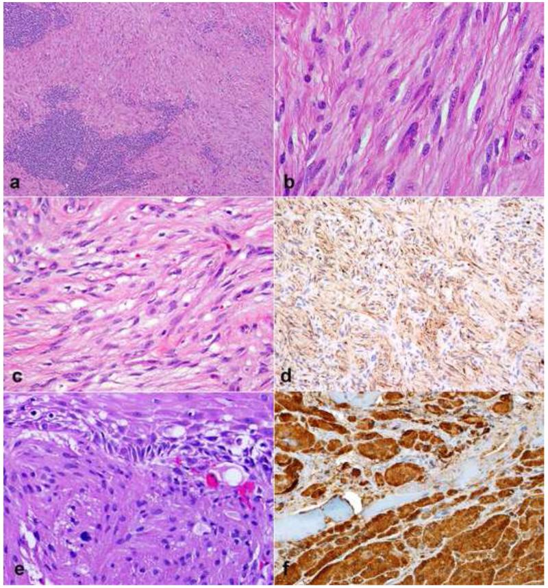
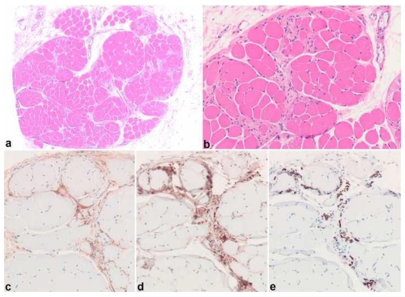
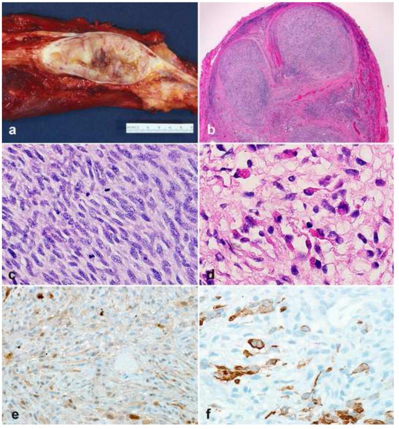
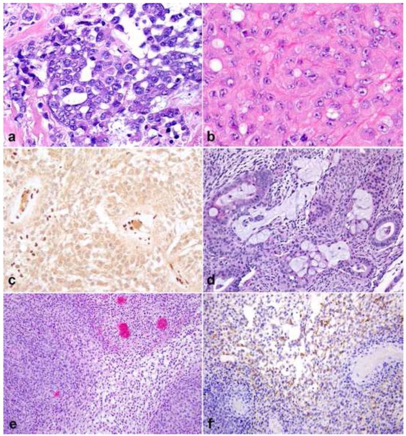
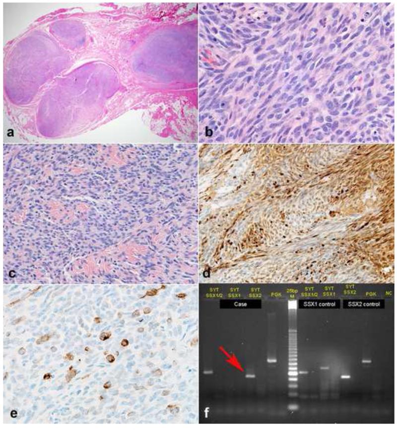
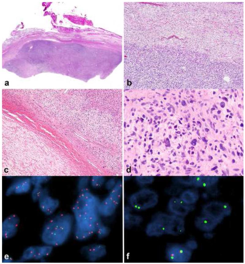
Peripheral nerve sheath tumors are common neoplasms, with classic identifiable features, but on occasion, they are diagnostically challenging. Although well-defined subtypes of peripheral nerve sheath tumors were described early in the history of surgical pathology, controversies regarding the classification and grading of these tumors persist. Advances in molecular biology have provided new insights into the nature of the various peripheral nerve sheath tumors, and have begun to suggest novel targeted therapeutic approaches. In this review, we discuss current concepts and problematic areas in the pathology of peripheral nerve sheath tumors. Diagnostic criteria and differential diagnosis for the major categories of nerve sheath tumors are proposed, including neurofibroma, schwannoma, and perineurioma. Diagnostically challenging variants, including plexiform, cellular and melanotic schwannomas are highlighted. A subset of these affects the childhood population, and has historically been interpreted as malignant, although current evidence and outcome data suggest they represent benign entities. The growing current literature and the author's experience with difficult to classify borderline or "hybrid tumors" are discussed and illustrated. Some of these classification gray zones occur with frequency in the gastrointestinal tract, an anatomical compartment that must always be entertained when examining these neoplasms. Other growing recent areas of interest include the heterogeneous group of pseudoneoplastic lesions involving peripheral nerve composed of mature adipose tissue and/or skeletal muscle, such as the enigmatic neuromuscular choristoma. Malignant peripheral nerve sheath tumors (MPNST) represent a diagnostically controversial group; difficulties in grading and guidelines to separate "atypical neurofibroma" from MPNST are provided. There is an increasing literature of MPNST mimics which neuropathologists must be aware of, including synovial sarcoma and ossifying fibromyxoid tumor. Finally, we discuss entities that are lacking from the section on cranial and paraspinal nerves in the current WHO classification, and that may warrant inclusion in future classifications. In summary, although the diagnosis and classification of most conventional peripheral nerve sheath tumors are relatively straightforward for the experienced observer, yet borderline and difficult-to-classify neoplasms continue to be problematic. In the current review, we attempt to provide some useful guidelines for the surgical neuropathologist to help navigate these persistent, challenging problems.


Figures

Figure 1
Figure 1. Pathologic Features of Neurofibroma
Neurofibromas may form circumscribed masses in dermis or soft tissue with variable, white-tan appearance (a). When involving a main peripheral nerve trunk, neurofibromas cause fusiform expansion (b). Wavy nuclei and “shredded carrot” type collagen are typical (c). Diffuse neurofibroma, a less common subtype, may form large masses with infiltration of fat and numerous pseudomeissnerian corpuscles (d). S100 usually labels a subset of cells of probable schwannian origin in neurofibromas (e), while CD34 shows variable reactivity, highlighting cells of an uncertain nature (f).
Figure 2
Figure 2. Diagnostically relevant neurofibroma variants
Plexiform neurofibromas typically form large, multinodular masses (a). Involvement and expansion of multiple peripheral nerve fascicles is definitional (b). Degenerative atypia, in the absence of hypercellularity has been interpreted by some authors as “atypical neurofibroma”, has no prognostic significance, but may create diagnostic difficulties (c). The atypia in some neurofibromas is reflected in hyperchromatic nuclei with “smudgy” chromatin, which is probably degenerative (d). Foci of hypercellularity may be present in some neurofibromas (e). Areas of hypercellularity (“cellular” neurofibromas) in the absence of other atypical features is still compatible with a benign course (f).
Figure 3
Figure 3. Rare neurofibroma changes and variants
Pigmented or melanotic change in neurofibroma is a very rare finding, usually encountered in the diffuse variant (a). Ossification may also be an occasional finding in neurofibromas (b). Massive soft tissue neurofibroma is a very rare neurofibroma variant essentially limited to NF1, and which may contain alarming, cellular areas (c).
Figure 4
Figure 4. Pathologic Features of Schwannoma
Schwannomas form well circumscribed, variegated masses with a tan appearance and macrocysts (a). Verocay bodies, characterized by distinct palisades with a fibrillary core are important diagnostic features (b). A well formed capsule is seen in almost all cases (c). Hyalinized vessels (d) are frequent findings. Diffuse immunoreactivity for S100 (e) and pericellular collagen IV (f) is almost universal in all schwannomas subtypes.
Figure 5
Figure 5. Important schwannoma variants
Cellular schwannomas, despite their alarming cellularity usually have a well formed capsule, often with multiple subcapsular lymphoid aggregates (a). Verocay bodies are usually lacking (b). Plexiform schwannomas involving major peripheral nerves are rare, characteristically involving multiple fascicles (c). Plexiform schwannomas are composed primarily of Antoni A areas (d). Mitotic activity in plexiform/cellular schwannomas may be brisk (arrows), a finding still compatible with a benign diagnosis(e). Electron microscopy demonstrates extensive basal lamina in all schwannoma subtypes, a diagnostically useful finding (f).
Figure 6
Figure 6. Rare schwannoma patterns and variants
Melanotic schwannoma is a unique variant that is difficult to separate from other melanocytic tumors (a). “Neuroblastoma-like” schwannoma is a rare variant characterized by collagenous rosettes surrounded by small cells with high nuclear cytoplasmic ratios (b). Pseudoglandular spaces mimicking glands may be rarely encountered (c). Epithelioid change in a schwannoma demonstrating otherwise reassuring features, including negligible proliferation and hyalinized vasculature (d).
Figure 7
Figure 7. Pathologic features of intraneural perineurioma
Intraneural perineurioma expands multiple nerve fascicles and may exhibit a multinodular gross appearance (photo courtesy of Dr. Robert Spinner)(a). An increase in perineurial and endoneurial cellularity is an important histologic clue (b). Immunohistochemical properties include EMA (c) and claudin (d) reactivity. S100 labels only residual Schwann cells (e), while neurofilament protein labels central axons within “pseudo-onion” bulbs (f)(arrows).
Figure 8
Figure 8. Benign nerve sheath tumor in NF1
Some benign nerve sheath tumors may be difficult to classify, even in clear cut genetic syndromes, containing areas typical of neurofibroma (a), or pattern variations reminiscent of schwannoma (b) and perineurioma (c).
Figure 9
Figure 9. Hybrid schwannoma-neurofibroma or “neurofibroma with Schwann cell nodules”
Nerve-associated intraorbital mass (a)(photo courtesy of Dr. James Garrity), with a plexiform architecture (b). Distinct schwannoma nodules and neurofibroma areas (c,d,e). Neurofilament protein immunostain highlights axons (f), while S100 strongly labels the schwannoma component (g).
Figure 10
Figure 10. Hybrid benign peripheral nerve sheath tumor
A subset of hybrid benign peripheral nerve sheath tumors present as soft tissue masses with prominent perineurial architecture (a). Areas of increased cellularity may be encountered (b). Strong S100 expression in a large number of cells reflects a Schwannian component (c). The perineurial component expresses EMA (d).
Figure 11
Figure 11. Gastrointestinal (GI) tract-associated benign nerve sheath tumors
The full range of benign nerve sheath tumors may involve the GI tract. Schwannomas at this site have variable features, which may include marked chronic inflammation (a) and non-specific spindle cell cytology that overlaps with GIST (b). Similarly, perineuriomas may show bland, non-specific spindle cell morphology (c), with specific diagnosis requiring immunohistochemistry for several markers, for example GLUT1 (d). Granular cell tumors represent another subset of neoplasms of presumed nerve sheath origin that favor the GI tract (e). Strong S100 expression is almost universal in these tumors (f).
Figure 12
Figure 12. Neuromuscular choristoma
Neuromuscular choristoma is an exquisitely rare pseudoneoplastic lesion of nerve characterized by intrafascicular replacement of nerve by mature skeletal muscle (a,b). Immunohistochemistry for EMA highlights the outlining perineurium (c), while S100 and neurofilament protein label associated Schwann cells (d) and axons (e).
Figure 13
Figure 13. Pathologic features of malignant peripheral nerve sheath tumor (MPNST)
MPNST create fleshy, variegated masses involving large peripheral nerve trunks (a). Multifascicular involvement is usually appreciated on cross sections (b). More often MPNST are characterized by uniform spindle cells with hyperchromatic nuclei arranged in fascicles (c). Heterologous elements, including myogenic differentiation may be present (d) Partial S100 immunoreactivity (e) and desmin expression (f) in a triton tumor.
Figure 14
Figure 14. Rare malignant peripheral nerve sheath tumor (MPNST) variants
Rare MPNSTs are composed of epithelioid cells that simulate carcinoma or melanoma (a). Rhabdoid cytology (b) and diffuse INI1 protein loss in neoplastic cells with preservation in vessels (c) may be present in rare cases, raising an important differential with epithelioid sarcoma. An even rarer variation in MPNST is overt epithelial differentiation in the form of well formed glands (d). Rare examples of MPNST may lack S100 expression almost entirely, and contain whorls (e) and EMA expression (f) suggestive of perineurial differentiation.
Figure 15
Figure 15. Intraneural synovial sarcoma mimicking malignant peripheral nerve sheath tumor (MPNST)
Synovial sarcoma represents the main entity in the differential diagnosis with MPNST, and similarly may infiltrate numerous nerve fascicles (a). Monotonous spindle cells in fascicles are characteristic (b) as well as variable intratumoral collagen bundles (c). Overt S100 immunoreactivity may be present in some synovial sarcomas (d). Cytokeratin typically labels isolated cells (e). Molecular confirmation may be required in some instances. In this example, RT-PCR reveals a SYT-SSX2 fusion transcript (arrow)(f), which is diagnostic of synovial sarcoma.
Figure 16
Figure 16. Low grade malignant peripheral nerve sheath tumor (MPNST)
Low grade MPNST represent diagnostically difficult problems but may be suspected by finding areas of hypercellularity within a conventional neurofibroma (a), in particular in the setting of NF1. A sharp interface between the precursor neurofibroma (upper) and low grade MPNST (lower) is usually evident (b). A collagenous interface may be seen in some cases (c). Minimal criteria for low grade MPNST require the presence of nuclear hyperchromasia, enlargement, and crowding (d). Molecular techniques such as fluorescence in situ hybridization (FISH) may be useful in a subset of cases, identifying genetic changes that are frequent in MPNST, including EGFR amplification (e) and homozygous CDKN2A deletions (f; a non-neoplastic cell at bottom serves as a positive internal control).

References

    1. Adamiak A, Lee CH, Nielsen TO, Webber D, O’Connell JX. Duodenal epithelioid gastrointestinal stromal tumor with prominent granular cell features. Hum Pathol. 2009;40:599–602. - PubMed
    1. Agaimy A, Wuensch PH. Perineurioma of the stomach. A rare spindle cell neoplasm that should be distinguished from gastrointestinal stromal tumor. Pathol Res Pract. 2005;201:463–467. - PubMed
    1. Agaimy A, Markl B, Kitz J, Wunsch PH, Arnholdt H, Fuzesi L, Hartmann A, Chetty R. Peripheral nerve sheath tumors of the gastrointestinal tract: a multicenter study of 58 patients including NF1-associated gastric schwannoma and unusual morphologic variants. Virchows Arch. 2010;456:411–422. - PubMed
    1. Agaram NP, Prakash S, Antonescu CR. Deep-seated plexiform schwannoma: a pathologic study of 16 cases and comparative analysis with the superficial variety. Am J Surg Pathol. 2005;29:1042–1048. - PubMed
    1. Ahrens WA, Ridenour RV, 3rd, Caron BL, Miller DV, Folpe AL. GLUT-1 expression in mesenchymal tumors: an immunohistochemical study of 247 soft tissue and bone neoplasms. Hum Pathol. 2008;39:1519–1526. - PubMed

MeSH terms

LinkOut - more resources