. 2012 Feb;24(2):428-43.
doi: 10.1105/tpc.111.093807. Epub 2012 Feb 10.

EARLY FLOWERING4 recruitment of EARLY FLOWERING3 in the nucleus sustains the Arabidopsis circadian clock

Affiliations

EARLY FLOWERING4 recruitment of EARLY FLOWERING3 in the nucleus sustains the Arabidopsis circadian clock

Eva Herrero et al. Plant Cell. 2012 Feb.

Abstract

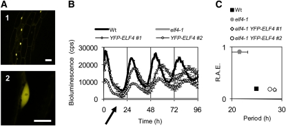
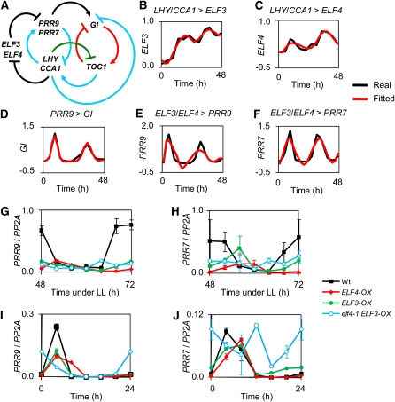
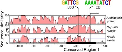
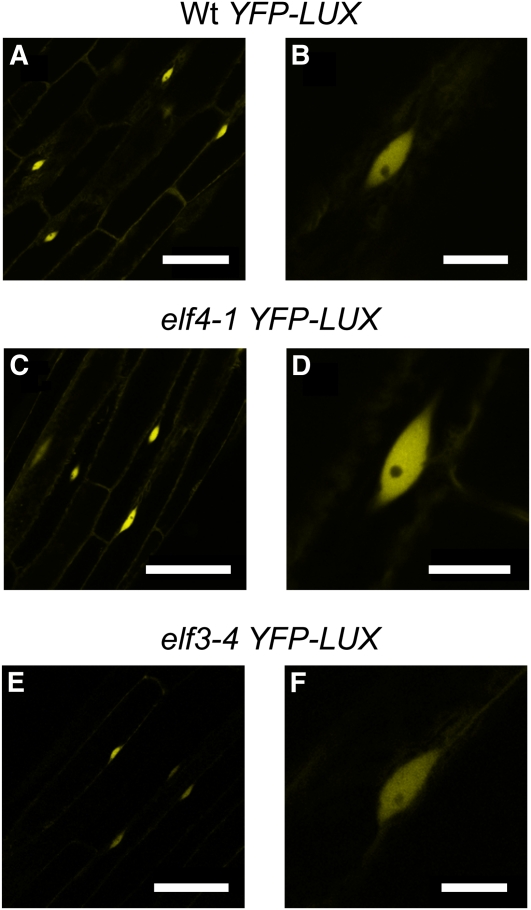
The plant circadian clock is proposed to be a network of several interconnected feedback loops, and loss of any component leads to changes in oscillator speed. We previously reported that Arabidopsis thaliana EARLY FLOWERING4 (ELF4) is required to sustain this oscillator and that the elf4 mutant is arrhythmic. This phenotype is shared with both elf3 and lux. Here, we show that overexpression of either ELF3 or LUX ARRHYTHMO (LUX) complements the elf4 mutant phenotype. Furthermore, ELF4 causes ELF3 to form foci in the nucleus. We used expression data to direct a mathematical position of ELF3 in the clock network. This revealed direct effects on the morning clock gene PRR9, and we determined association of ELF3 to a conserved region of the PRR9 promoter. A cis-element in this region was suggestive of ELF3 recruitment by the transcription factor LUX, consistent with both ELF3 and LUX acting genetically downstream of ELF4. Taken together, using integrated approaches, we identified ELF4/ELF3 together with LUX to be pivotal for sustenance of plant circadian rhythms.


Figures

Figure 1.
Figure 1.
ELF3 Is Downstream of ELF4. (A) and (B) CCA1:LUC under LD (12 h light/12 h dark). CCA1:LUC expression was severely reduced in elf3-4, elf4-1, and elf3-4 elf4-1 compared with the wild type (Wt). Note the different resolutions of the y axes and the lack of driven CCA1:LUC oscillations in elf3-4 and elf3-4 elf4-1. (C) and (D) Bioluminescence of LHY:LUC under LL. Period length ± sd: (C) the wild type, 25.1 ± 1.3 h; ELF4-OX, 28.75 ± 0.95 h; (D) the wild type, 25.1 ± 1.3 h; ELF3-OX, 25.9 ± 1.6 h; elf4-1 ELF3-OX, 25.2 ± 0.9 h. Note that in elf3-4 and elf3-4 ELF4-OX (C), and in elf4-1 (D), LHY:LUC is nearly undetectable (arrow), but elf4-1 ELF3-OX regains LHY:LUC rhythms (D). The wild type is the same in both panels. Error bars indicate se, n = 24. cps, count per second. This experiment is representative of at least three independent replicates. (E) Bioluminescence of CCR2:LUC under LL. Period length estimates: the wild type, 27.4 ± 0.9 h; ELF3-OX, 27.8 ± 1.1 h; elf4-1 ELF3-OX, 28.9 ± 0.8 h. Error bars indicate se. (F) Bioluminescence of CCR2:LUC in DD. Period length estimates: the wild type, 28.0 ± 1.3 h; ELF3-OX, 29.5 ± 1.7 h; elf4-1 ELF3-OX, 30.3 ± 1.4 h. Error bars indicate se.
Figure 2.
Figure 2.
The Middle Domain of ELF3 Mediates the Physical Interaction with ELF4. (A) The middle domain of ELF3 (ELF3M; residues 261 to 484) mediates the physical interaction with ELF4. Y2H assay of DB-ELF4 and ELF3-AD fragments. AD-ELF3 fragments are defined in Figure 3A. empty, AD or DB only; –LW and –LWH, dropout for Leu/Trp and Leu/Trp/His, respectively; 3AT, 3-amino-1,2,4-triazole. The Y2H experiments were performed three times with similar results. (B) ELF3 and ELF4 interact in planta. FRET assay of ELF3-CFP and YFP-ELF4 in N. benthamiana (using the 35S promoter), n = 10. Error bars indicate se. This experiment is representative of three independent replicates. (C) ELF4 binds to the ELF3 middle domain (ELF3M) in vitro. SDS-PAGE gel: lane 1, fraction of His6-MBP-ELF3M fragment eluted from the amylose column; lane 2, fraction of His6-ELF4 eluted from the Ni-nitriloacetic acid column; lane 3, fraction of His6-ELF4 fragment eluted from the amylose column (no ELF4 band, indicating lack of affinity for amylose); lane 4, ELF4 pulldown in the ELF3M:ELF3M-ELF4 fraction eluted from amylose resin, followed by size exclusion chromatography. Asterisk indicates contamination from proteolysis of MBP-ELF3M. MW, molecular weight.
Figure 3.
Figure 3.
ELF4 Increases ELF3 Abundance in the Nucleus. (A1) ELF3 fragments used for Y2H with the ELF3 AD (Figure 1) and for the ELF3 YFP constructs. The ELF4 arrow pointing to ELF3M represents the protein–protein interaction. (A1) to (A7) Color bars: ELF3F (full length, black), ELF3N (residues 1 to 259, pink), ELF3M (residues 261 to 484, green), ELF3C (residues 485 to 695, blue), and ELF4 (full length, gray). (A2) to (A7) YFP channel of epidermal cells of N. benthamiana infiltrated with YFP-ELF3 fragments. The color code refers to the scheme to the left. Bars = 20 μm. (A2) YFP-ELF3F, magnification highlighted with an asterisk. (A3) YFP-ELF3N is cytoplasmic. (A4) and (A5) YFP-ELF3NM and YFP-ELF3M have both a cytoplasmic and nuclear distribution. (A6) and (A7) YFP-ELF3MC and YFP-ELF3C are both localized in the nucleus. (A8) CFP channel of epidermal cells of N. benthamiana infiltrated with ELF4-CFP. The photos are representative of three independent experiments. (B1) to (B5) ELF4 increases ELF3M nuclear localization. Expression of YFP-ELF3M only: YFP channel (B1). Coexpression of YFP-ELF3M and ELF4-CFP: YFP (B2) and CFP (B3) channel. Signal intensity of YFP channel: YFP-ELF3 alone (B4) from experiment in (B1); coexpression of YFP-ELF3M and ELF4-CFP (B5) from the experiment in (B2). The z, x, and y axes represent YFP intensity, horizontal plane, and vertical plane, respectively. Black arrows in (B1) and (B2) correspond to white arrows in (B4) and (B5). This experiment is representative of three independent replicates.
Figure 4.
Figure 4.
ELF3 Localizes in Nuclear Bodies in the Presence of ELF4. Transient expression of ELF3 and ELF4 YFP/CFP fusion proteins in N. benthamiana epidermal cells. (A1) and (A2) Individual expression of ELF3pro:ELF3-YFP ([A1]; YFP channel) and ELF4p:CFP-ELF4 ([A2]; CFP channel). The images represent the average of the 15-μm z-stack (10 slices). Asterisks indicate the nuclei magnified in bottom right corner in (A1) and (A2). Bars = 20μm. (B1) and (B2) Coexpression of ELF3pro:ELF3-YFP and ELF4pro:ELF4-CFP. YFP channel (B1); CFP channel (B2). The images represent the average of the 15-μm z-stack (10 slices). Bars = 20 μm. (C1) to (C3) Nucleus where ELF3pro:ELF3-YFP and ELF4pro:ELF4-CFP are coexpressed. YFP channel (C1), CFP channel (C2), and merged YFP-CFP (C3). Bars = 5 μm.
Figure 5.
Figure 5.
Subcellular Distribution of ELF3 in Arabidopsis Stable Transgenic Lines. Confocal microscopy of Arabidopsis expressing YFP-ELF3 fragments (under the 35S promoter). (1) YFP-ELF3, (2) YFP-ELF3N, (3) YFP-ELF3NM, (4) YFP-ELF3M, (5) YFP-ELF3MC, (6) YFP-ELF3C, (7) YFP-ELF3F nuclei, (8) YFP-ELF3MC nuclei, and (9) YFP-ELF3C nuclei. Maximum projection of 6-μm stacks (1 to 6) and 4-μm stacks for nuclei (7 to 9). Bars = 50 μm in (1) to (6) and 10 μm in (7) to (9). The imaging was repeated two times with similar results.
Figure 6.
Figure 6.
Defining a Functional Region of ELF3. (A) to (C) Circadian rhythmicity of LHY:LUC under LL. Wild-type trace is the same in (A) and (B). Two representative single-insertion lines for each of the YFP transgenes are shown (indicated by #1 and #2, respectively). (A) YFP-ELF3 overexpression restores circadian oscillations and causes a lengthening of the period in the elf3-4 background. Arrow indicates the low activity of LHY:LUC in elf3-4. (B) YFP-ELF3MC overexpression restores elf3-4 circadian oscillations and causes a lengthening of the period and low amplitude. Arrow indicates the elf3-4 trace. Wild-type trace is the same in (A) and (B). Error bars indicate se, n = 24. (C) Period versus relative amplitude error (RAE) of LHY:LUC under LL in (A) and (B). Period and relative amplitude error estimates ± se: the wild type, 25.4 ± 0.2 h/0.2 ± 0.01; elf3-4, 24.1 ± 0.9 h/0.82 ± 0.04; YFP-ELF3 #1, 27.6 ± 0.3 h/0.21 ± 0.04; YFP-ELF3 #2, 27.1 ± 0.3 h/0.26 ± 0.02; YFP-ELF3MC #1, 26.8 ± 0.4 h/0.17 ± 0.01; YFP-ELF3MC #2, 26.6 ± 0.3 h/0.2 ± 0.01.
Figure 7.
Figure 7.
Subcellular Localization of ELF4 in Arabidopsis Stable Transgenic Lines. (A) Confocal microscopy of hypocotyl cells from stable Arabidopsis lines expressing YFP-ELF4 under the 35S promoter in the elf4-1 background. Bars = 25 μm in (1) and 10 μm in (2). The images are representative of at least two independent elf4-1 YFP-ELF3-OX lines (indicated as #1 and #2, respectively). The imaging was performed twice with similar results. (B) Normalized bioluminescence of LHY:LUC under LL for the wild type (Wt), elf4-1, and elf4-1 YFP-ELF4 lines. Arrow indicates the low activity of LHY:LUC in elf4-1. cps, counts per second. (C) Period versus relative amplitude error (R.A.E.) plot of data from panel (B). Period and relative amplitude error values ± se: the wild type, 24.93 ± 0.2 h/0.2 ± 0.01; elf4-1, 21.50 ± 0.17 h/0.91 ± 0.03; elf4-1 YFP-ELF4 #1, 28.63 ± 0.24 h/0.18 ± 0.01; elf4-1 YFP-ELF4 #2, 27.61 ± 0.19 h/0.19 ± 0.01. Error bars indicate se, n = 24. This experiment was performed at least twice with similar results.
Figure 8.
Figure 8.
LTI Modeling Includes ELF3/ELF4 in the Circadian Network. (A) Expanded circadian clock model incorporates ELF3 and ELF4. LTI models of the circadian system using a data set of clock gene expression data from LD and LL (Kolmos et al., 2011) as training data for the systems identification. Newly described links are in black. Links common between the three-loop model (Locke et al., 2006) and our model are blue (>60% fitness) and green (50 to 60% fitness). Links existing in the three-loop model, but not in our model, are in red. (B) to (F) Fitness of LTI models to real expression data. Real (black) and simulated (red) data curves of gene expression: LHY/CCA1 links to ELF3 (B), LHY/CCA1 links to ELF4 (C), PRR9 links to GI (D), ELF3/ELF4 links to PRR9 (E), and ELF3/ELF4 links to PRR7 (F). (G) and (H) PRR9 (G) and PRR7 (H) transcript accumulation under LL in the wild type, ELF3-OX, ELF4-OX, and elf4-1 ELF3-OX. Samples were collected after 48 h under LL. Expression values are normalized to PP2A and are representative of two biological replicates. (I) and (J) PRR9 (I) and PRR7 (J) transcript accumulation under LD (short days, 8 h light/16 dark) in the wild type, ELF3-OX, ELF4-OX, and elf4-1 ELF3-OX. Expression values are normalized to PP2A and are representative of two biological replicates. Wt, wild type.
Figure 9.
Figure 9.
Phylogenetically Conserved Region of the PRR9 Promoter. Pairwise alignments of the A. thaliana PRR9 promoter to orthologous sequences of A. lyrata, C. rubella, and A. alpina, respectively, shown as VISTA plots. Light-red color indicates regions where a sliding window of at least 30 bp has >70% identity. Conserved Region 1 (−331 to +89) is highlighted with a black line. Vertical bars indicate the position of highly conserved LBS, EE, and the translational initiation codon (arrow). Conservation of LBS and EE is shown as WEBLOGO.
Figure 10.
Figure 10.
LUX Nuclear Localization Is Independent of ELF4 and ELF3. YFP-LUX nuclear localization is not affected in the elf4-1 and elf3-4 backgrounds. YFP confocal microscopy of hypocotyl epidermal cells from Arabidopsis YFP-LUX lines. The wild type ([A] and [B]), elf4-1 ([C] and [D]), and elf3-4 ([E] and [F]). Bars = 50 μm in (A), (C), and (E) and 10 μm in (B), (D), and (F). The images are representative of two independent lines per background. The experiments were performed twice with similar results.
Figure 11.
Figure 11.
LUX Is Downstream of ELF4 Action. (A) to (C) LHY:LUC rhythms under LL of YFP-LUX in the wild type (A), elf4-1 (B), and elf3-4 (C) background, respectively. Two representative single-insertion lines for each of the YFP transgenes are shown (indicated by #1 and #2). Error bars indicate se, n = 24. Arrows indicate low levels of LHY:LUC in elf4-1 (B) and elf3-4 (C). (D) Period versus relative amplitude error (RAE) of LHY:LUC rhythms from data in (A) to (C). Period length/relative amplitude error ± se: the wild type, 27.7 ± 0.4 h/0.28 ± 0.05; elf3-4, 23.7 ± 3.9 h/0.94 ± 0.03; elf4-1, 27.9 ± 2.8 h/0.96 ± 0.03; wild-type YFP-LUX #1, 26.8 ± 0.8 h/0.36 ± 0.04; wild-type YFP-LUX #2, 27.3 ± 1.1 h/0.35 ± 0.05; elf3-4 YFP-LUX #1, 24.2 ± 3.1 h/0.96 ± 0.02; elf3-4 YFP-LUX #2, 28.5 ± 2.6 h/0.93 ± 0.03; elf4-1 YFP-LUX #1, 26.3 ± 0.7 h/0.38 ± 0.04; elf4-1 YFP-LUX #2, 26.4 ± 0.6 h/0.32 ± 0.05.

References

    1. Alabadí D., Oyama T., Yanovsky M.J., Harmon F.G., Más P., Kay S.A. (2001). Reciprocal regulation between TOC1 and LHY/CCA1 within the Arabidopsis circadian clock. Science 293: 880–883 - PubMed
    1. Brudno M., Malde S., Poliakov A., Do C.B., Couronne O., Dubchak I., Batzoglou S. (2003). Glocal alignment: finding rearrangements during alignment. Bioinformatics 19(suppl. 1): i54–i62 - PubMed
    1. Chen M. (2008). Phytochrome nuclear body: An emerging model to study interphase nuclear dynamics and signaling. Curr. Opin. Plant Biol. 11: 503–508 - PubMed
    1. Chen M., Galvão R.M., Li M., Burger B., Bugea J., Bolado J., Chory J. (2010). Arabidopsis HEMERA/pTAC12 initiates photomorphogenesis by phytochromes. Cell 141: 1230–1240 - PMC - PubMed
    1. Covington M.F., Maloof J.N., Straume M., Kay S.A., Harmer S.L. (2008). Global transcriptome analysis reveals circadian regulation of key pathways in plant growth and development. Genome Biol. 9: R130. - PMC - PubMed

Publication types

MeSH terms

Substances

Associated data