. 2012 May;79(1-2):21-33.
doi: 10.1007/s11103-012-9891-4. Epub 2012 Feb 11.

Unraveling the function of Arabidopsis thaliana OS9 in the endoplasmic reticulum-associated degradation of glycoproteins

Affiliations

Unraveling the function of Arabidopsis thaliana OS9 in the endoplasmic reticulum-associated degradation of glycoproteins

Silvia Hüttner et al. Plant Mol Biol. 2012 May.

Abstract

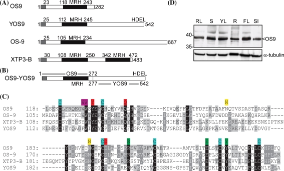
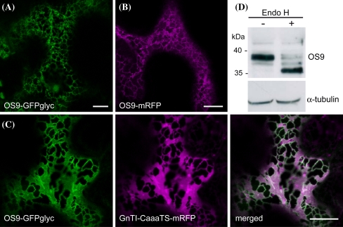
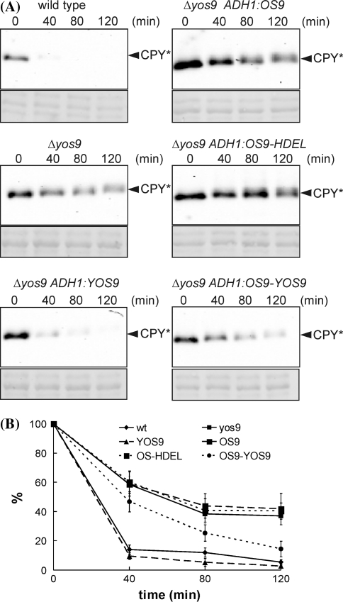
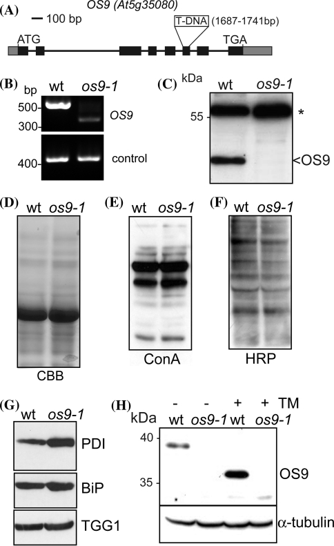
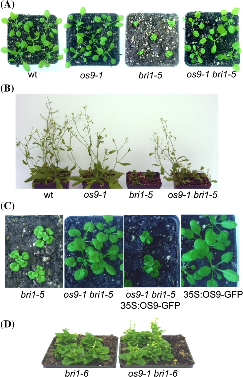
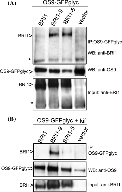
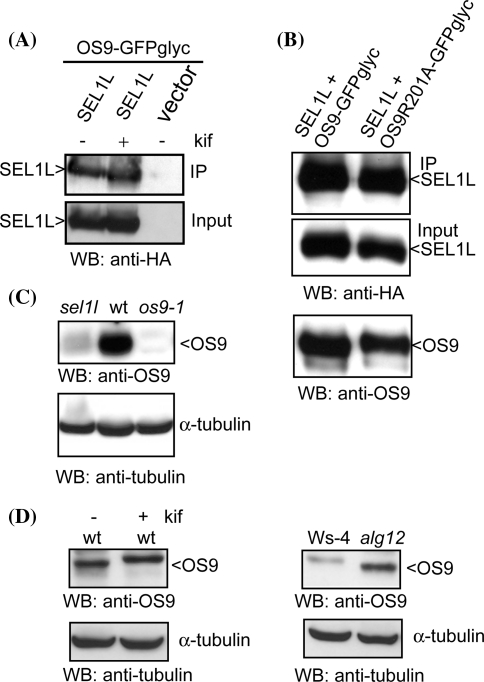
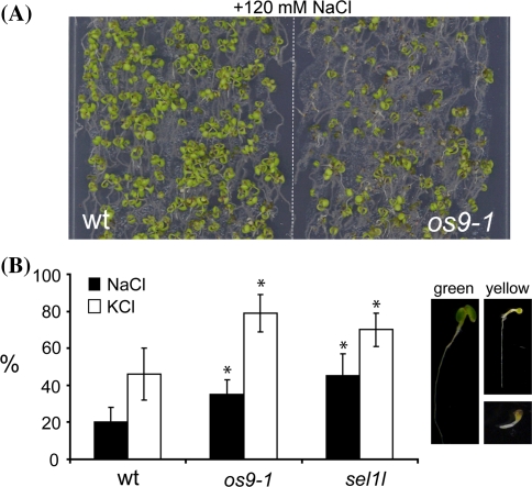
In the endoplasmic reticulum, immature polypeptides coincide with terminally misfolded proteins. Consequently, cells need a well-balanced quality control system, which decides about the fate of individual proteins and maintains protein homeostasis. Misfolded and unassembled proteins are sent for destruction via the endoplasmic reticulum-associated degradation (ERAD) machinery to prevent the accumulation of potentially toxic protein aggregates. Here, we report the identification of Arabidopsis thaliana OS9 as a component of the plant ERAD pathway. OS9 is an ER-resident glycoprotein containing a mannose-6-phosphate receptor homology domain, which is also found in yeast and mammalian lectins involved in ERAD. OS9 fused to the C-terminal domain of YOS9 can complement the ERAD defect of the corresponding yeast Δyos9 mutant. An A. thaliana OS9 loss-of-function line suppresses the severe growth phenotype of the bri1-5 and bri1-9 mutant plants, which harbour mutated forms of the brassinosteroid receptor BRI1. Co-immunoprecipitation studies demonstrated that OS9 associates with Arabidopsis SEL1L/HRD3, which is part of the plant ERAD complex and with the ERAD substrates BRI1-5 and BRI1-9, but only the binding to BRI1-5 occurs in a glycan-dependent way. OS9-deficiency results in activation of the unfolded protein response and reduces salt tolerance, highlighting the role of OS9 during ER stress. We propose that OS9 is a component of the plant ERAD machinery and may act specifically in the glycoprotein degradation pathway.


Figures

Fig. 1
Fig. 1
OS9 is a MRH domain protein that is ubiquitously expressed. a A comparison of the domain organization of OS9 with yeast (YOS9) and human (OS-9/XTP3-B) MRH domain proteins is shown. The signal peptide is indicated in grey, the MRH domain is shown in black. For OS-9 and XTP3-B the longest variants are shown (Hosokawa et al. 2010). b The chimeric OS9-YOS9 protein that was used for complementation of Δyos9 yeast consists of amino acids 1–272 from Arabidopsis OS9 fused to amino acids 277–542 from yeast YOS9. c Sequence alignment of MRH domains was performed using ClustalW (http://www.ebi.ac.uk/Tools/clustalw2/index.html). The domains were identified according to Munro (2001). Conserved residues are shaded (100% identity black, 80% identity dark grey, 60% identity light grey). The six conserved cysteines involved in the formation of disulphide bonds are marked in blue, amino acid residues involved in carbohydrate binding are marked in red (Roberts et al. ; Szathmary et al. 2005) and putative N-glycosylation sites are marked in yellow. The two tryptophan residues that determine the α1,6-mannose binding specificity of human OS-9 are marked in magenta (Satoh et al. 2010) and the three mannose binding residues of YOS9 are marked in green (Ghosh et al. ; Bhamidipati et al. 2005). d Proteins were extracted from different A. thaliana organs or developmental stages (RL rosette leaves, S whole seedlings, YL young leaves, R root, FL flower, SI siliques), separated by SDS–PAGE and analyzed by immunoblots using OS9-specific antibodies. α-tubulin expression was used as a control
Fig. 2
Fig. 2
Fluorescent protein-tagged OS9 displays ER localization. Confocal microscopy of N. benthamiana leaf epidermal cells expressing a OS9-GFPglyc, b OS9-mRFP and c OS9-GFPglyc (in green) together with the ER-retained construct GnTI-CaaaTS-mRFP (in magenta) reveals ER localization of OS9 (merged image). Scale bars = 10 μM. d Immunoblot analysis of the glycosylation status of endogenous OS9. Samples were subjected to Endo H digestion, separated by SDS–PAGE and analyzed by immunoblotting with OS9-specific antibodies or α-tubulin as a control
Fig. 3
Fig. 3
A chimeric OS9-YOS9 protein suppresses the yeast Δyos9 defect. a Equal cell numbers of yeast strains expressing the ERAD substrate CPY* and OS9 or YOS9 were incubated with cycloheximide and proteins were extracted at the given time points. Shown are representative immunoblot images. The lower panel shows a Ponceau S staining of the membrane. b Signal intensities (vertical axis) of the CPY* specific band from three independent experiments were quantified and blotted against time. The amount of the CPY* protein band at time point 0 was set to 100%
Fig. 4
Fig. 4
os9-1 lacks a functional OS9 protein and displays activation of the UPR. a Schematic overview of the OS9 gene structure. Boxes represent exons (the black area represents the coding region), the os9-1 T-DNA insertion is indicated. b Reverse transcription-PCR analysis of the os9-1 mutant using oligonucleotides that flank the insertion site. UBQ5 amplification served as a control. c Immunoblot with anti-OS9 antibodies. The asterisk indicates an unspecific band, which was used as a loading control. d Protein extracts from wt and os9-1 14-day-old seedlings were subjected to SDS–PAGE and Coomassie brilliant blue (CBB) staining. e Blots were analyzed using concanavalin A (ConA) (f) or anti-horseradish peroxidase (HRP) antibodies, which recognize N-glycans with β1,2-xylose and core α1,3-fucose residues. g Protein extracts were analyzed by immunoblotting using anti-PDI, anti-BiP2 and anti-TGG1 antibodies, respectively. h 10-day-old seedlings were incubated for 24 h in ½× MS medium supplemented with 3% sucrose and 5 μg mL−1 tunicamycin (TM), protein extracts were analyzed using OS9 or α-tubulin antibodies
Fig. 5
Fig. 5
os9-1 suppresses the bri1-5 phenotype by affecting the ER-retention of BRI1-5. a 17-day and b 4-week-old soil-grown plants. c Expression of OS9-GFP in os9-1 bri1-5 plants restores the bri1-5 growth phenotype. os9-1 bri1-5 double mutants were floral dipped with 35S:OS9-GFP. Expression of 35S:OS9-GFP in wild-type is shown as a control. d 10-week-old bri1-6 single and os9-1 bri1-6 double mutants display the bri1-6 dwarf phenotype
Fig. 6
Fig. 6
OS9 interacts with the ERAD substrates BRI1-5 and BRI1-9. a BRI1 forms were transiently co-expressed with OS9-GFPglyc in N. benthamiana leaves. OS9-GFPglyc was purified and co-purified protein fractions were analyzed by SDS–PAGE and immunoblotting with anti-BRI1 and anti-OS9 antibodies. The asterisk indicates an unspecific band that was used as loading control. b As in a but expression was done in the presence of 20 μM kifunensine (kif)
Fig. 7
Fig. 7
OS9 interacts with SEL1L in a glycan-independent way. a HA-tagged A. thaliana SEL1L was co-expressed with OS9-GFPglyc in N. benthamiana. OS9-GFPglyc was purified and co-purified protein fractions were analyzed by immunoblotting with anti-HA antibodies. (+) and (−) indicates expression in the presence or absence of 20 μM kifunensine (kif). b As in a but the mutated OS9R201A-GFPglyc was used for purification of SEL1L. c Protein extracts from A. thaliana sel1l, wild-type (wt) and os9-1 were separated by SDS–PAGE and analyzed by immunoblotting with anti-OS9 antibodies. α-tubulin expression was used as a control. d As in c but wt with kif and alg12 are shown. alg12 is in Ws-4 background. OS9 is more abundant in alg12 than in Ws-4, which is very likely caused by the activation of the unfolded protein response in alg12 (Hong et al. 2009)
Fig. 8
Fig. 8
os9-1 seedlings are sensitive to salt stress. a Wild-type (wt) and os9-1 seedlings were directly spread on ½× MS medium containing 1.5% sucrose supplemented with 120 mM NaCl and grown for 12 days. b Quantitative analysis of different seedling phenotypes grown for 12 days either on 120 mM NaCl or KCl. Percentages (vertical axis) represent smaller and yellow seedlings and are means ± SE from three independent repeats (more than 100 seedlings were counted per line and experiment, the total number of seedlings represents 100%). *P < 0.05 (paired Student’s t test)

Similar articles

Cited by

References

    1. Bernasconi R, Galli C, Calanca V, Nakajima T, Molinari M. Stringent requirement for HRD1, SEL1L, and OS-9/XTP3-B for disposal of ERAD-LS substrates. J Cell Biol. 2010;188:223–235. doi: 10.1083/jcb.200910042. - DOI - PMC - PubMed
    1. Bhamidipati A, Denic V, Quan EM, Weissman JS. Exploration of the topological requirements of ERAD identifies Yos9p as a lectin sensor of misfolded glycoproteins in the ER lumen. Mol Cell. 2005;19:741–751. doi: 10.1016/j.molcel.2005.07.027. - DOI - PubMed
    1. Brandizzi F, Hanton S, DaSilva L, Boevink P, Evans D, Oparka K, Denecke J, Hawes C. ER quality control can lead to retrograde transport from the ER lumen to the cytosol and the nucleoplasm in plants. Plant J. 2003;34:269–281. doi: 10.1046/j.1365-313X.2003.01728.x. - DOI - PubMed
    1. Ceriotti A. Waste disposal in the endoplasmic reticulum, ROS production and plant salt stress response. Cell Res. 2011;21:555–557. doi: 10.1038/cr.2011.18. - DOI - PMC - PubMed
    1. Christianson JC, Shaler TA, Tyler RE, Kopito RR. OS-9 and GRP94 deliver mutant α1-antitrypsin to the Hrd1-SEL1L ubiquitin ligase complex for ERAD. Nat Cell Biol. 2008;10:272–282. doi: 10.1038/ncb1689. - DOI - PMC - PubMed

Publication types

MeSH terms