. 2012 Apr 15;302(8):H1726-36.
doi: 10.1152/ajpheart.00783.2011. Epub 2012 Feb 10.

Testosterone and interleukin-1β increase cardiac remodeling during coxsackievirus B3 myocarditis via serpin A 3n

Affiliations

Testosterone and interleukin-1β increase cardiac remodeling during coxsackievirus B3 myocarditis via serpin A 3n

Michael J Coronado et al. Am J Physiol Heart Circ Physiol. .

Abstract

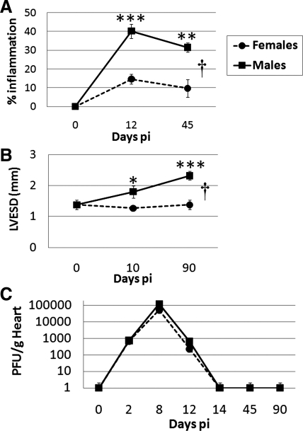
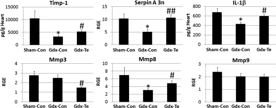
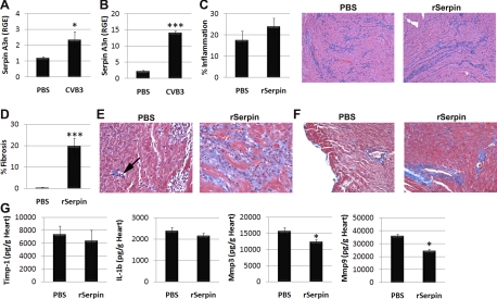
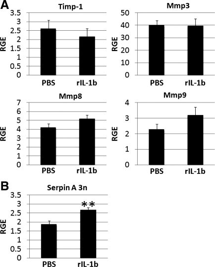
Myocarditis and dilated cardiomyopathy (DCM) are often caused by viral infections and occur more frequently in men than in women, but the reasons for the sex difference remain unclear. The aim of this study was to assess whether gene changes in the heart during coxsackievirus B3 (CVB3) myocarditis in male and female BALB/c mice predicted worse DCM in males. Although myocarditis (P = 4.2 × 10(-5)) and cardiac dilation (P = 0.008) were worse in males, there was no difference in viral replication in the heart. Fibrotic remodeling genes, such as tissue inhibitor of metalloproteinase (TIMP)-1 and serpin A 3n, were upregulated in males during myocarditis rather than during DCM. Using gonadectomy and testosterone replacement, we showed that testosterone increased cardiac TIMP-1 (P = 0.04), serpin A 3n (P = 0.007), and matrix metalloproteinase (MMP)-8 (P = 0.04) during myocarditis. Testosterone increased IL-1β levels in the heart (P = 0.02), a cytokine known to regulate cardiovascular remodeling, and IL-1β in turn increased cardiac serpin A 3n mRNA (P = 0.005). We found that 39 of 118 (33%) genes identified in acute DCM patients were significantly altered in the heart during CVB3 myocarditis in mice, including serpin A 3n (3.3-fold change, P = 0.0001). Recombinant serpin A 3n treatment induced cardiac fibrosis during CVB3 myocarditis (P = 0.0008) while decreasing MMP-3 (P = 0.04) and MMP-9 (P = 0.03) levels in the heart. Thus, serpin A 3n was identified as a gene associated with fibrotic cardiac remodeling and progression to DCM in male myocarditis patients and mice.


Figures

Fig. 1.
Fig. 1.
Sex differences in coxsackievirus B3 (CVB3) myocarditis in relation to cardiac function and viral replication. Male and female BALB/c mice were inoculated intraperitoneally with 103 plaque-forming units (PFU) of heart-passaged CVB3 on day 0, and acute [days 10 or 12 postinfection (pi)] and chronic (days 45 or 90 postinfection) myocarditis (percent inflammation) and cardiac function [left ventricular end-systolic dimension (LVESD)] were assessed by histology (A) or echocardiography (B), respectively. C: viral replication in the heart was examined at various time points postinfection using a plaque assay. Student's t-test compared females with males at individual time points: *P < 0.05; **P < 0.01; ***P < 0.001. ANOVA compared females with males over time: †P = 4.12 × 10⁻⁵ in A and †P = 0.008 in B. Data show means ± SE of 7–10 mice/group.
Fig. 2.
Fig. 2.
Cardiovascular remodeling genes increase during acute CVB3 myocarditis in male mice compared with undiseased control males. Male BALB/c mice were inoculated intraperitoneally with sterile PBS or CVB3 on day 0, and microarrays were conducted on individual hearts (n = 3 hearts/group) on day 10 postinfection (A; acute myocarditis) or day 90 postinfection (B; chronic myocarditis/dilated cardiomyopathy). Ingenuity pathway analysis of microarray data revealed that a major gene network associated with acute CVB3 myocarditis was “cardiovascular disease, organismal injury and abnormality, and tissue morphology.” Red/pink and green represent genes significantly up- or downregulated during acute or chronic myocarditis compared with undiseased controls, respectively.
Fig. 3.
Fig. 3.
Testosterone increases tissue inhibitor of metalloproteinase (Timp1), serpin A 3n, IL-1β, and matrix metalloproteinase (Mmp8) in the heart of male mice during acute CVB3 myocarditis. Male BALB/c mice received a sham operation and a control capsule without testosterone (Sham-Con mice), were gonadectomized and received a control capsule (Gdx-Con mice), or gonadectomized and received a testosterone replacement capsule (Gdx-Te mice). After recovering from the gonadectomy for 2 wk, mice received CVB3 on day 0, and the expression of cardiac remodeling genes was examined at day 10 postinfection by ELISA (pg/ml) or RT-PCR [relative gene expression (RGE)]. Data show means ± SE; n = 10 mice/group. *Gdx-Con or Gdx-Te mice compared with Sham-Con mice by Student's t-test; #Gdx-Te mice compared with Gdx-Con mice by Student's t-test; * or #P < 0.05; ##P < 0.01. The three groups were significantly different by two-way ANOVA for Timp1, serpin A 3n, and IL-1β (†P < 0.05).
Fig. 4.
Fig. 4.
Recombinant serpin A 3n (rSerpin) promotes cardiac remodeling during CVB3 myocarditis. Male BALB/c mice received sterile PBS or CVB3 intraperitoneally on day 0, and serpin A 3n levels were examined in the heart by RT-PCR on day 2 (A) or day 10 (B) postinfection. Male BALB/c mice were inoculated intraperitoneally with CVB3 on day 0, and either sterile PBS or rSerpin (10 μg/ml) was injected intraperitoneally every other day from day 1 to day 9 postinfection. Mice were examined at day 10 postinfection. C: summary of histology score (left) and representative hematoxylin and eosin staining for inflammation (purple; right). Magnification: ×64. D: percent fibrosis was calculated as the area with collagen deposition compared with the entire myocardial section using a microscope eyepiece grid. Masson's trichrome to detect collagen deposition (bright blue) showed areas of inflammation (E) or perivascular fibrosis (F). Magnification: ×250 in E and ×100 in F. E: small area of fibrosis (arrow) compared with nonfibrotic inflammation in PBS mice (left) versus widespread fibrosis associated with inflammation in rSerpin-treated mice (right). Data show means ± SE of 10 mice/group in A–C, D, and G. *P < 0.05; ***P < 0.001.
Fig. 5.
Fig. 5.
Recombinant IL-1β (rIL-1b) increases serpin A 3n levels in the heart of males during CVB3 myocarditis. A and B: cardiac remodeling genes were assessed by RT-PCR during acute myocarditis (day 10 postinfection) in male BALB/c mice that had been treated with recombinant IL-1β from day 1 to day 9 postinfection (n = 10 mice/group). RGE, normalized to hypoxanthine phosphoribosyltransferase (HPRT), is shown as means ± SE of 10 mice/group. **P < 0.01.
Fig. 6.
Fig. 6.
Influence of testosterone (Te) on cardiac remodeling. Testosterone increases IL-1β and serpin A 3n levels in the heart during acute CVB3 myocarditis. IL-1β further elevates serpin A 3n levels, which contribute to cardiac remodeling and fibrosis by reducing Mmp3 and Mmp9.

References

    1. Asakura M, Kitakaze M. Global gene expression profiling in the failing myocardium. Circ J 73: 1568– 1576, 2009 - PubMed
    1. Barth AS, Kuner R, Buness A, Ruschhaupt M, Merk S, Zwermann L, Kääb S, Kreuzer E, Steinbeck G, Mansmann U, Poustka A, Nabauer M, Sültmann H. Identification of a common gene expression signature in dilated cardiomyopathy across independent microarray studies. J Am Coll Cardiol 48: 1610– 1617, 2006 - PubMed
    1. Blauwet LA, Cooper LT. Myocarditis. Prog Cardiovasc Dis 52: 274– 288, 2010 - PMC - PubMed
    1. Brauer PR. MMPs–role in cardiovascular development and disease. Front Biosci 11: 447– 478, 2006 - PubMed
    1. Caforio ALP, Calabrese F, Angelini A, Tona F, Vinci A, Bottaro S, Ramondo A, Carturan E, Iliceto S, Thiene G, Daliento L. A prospective study of biopsy-proven myocarditis: prognostic relevance of clinical and aetiopathogenetic features at diagnosis. Eur Heart J 28: 1326– 1333, 2007 - PubMed

Publication types

MeSH terms