. 2012 May 1;125(Pt 9):2172-84.
doi: 10.1242/jcs.096214. Epub 2012 Feb 10.

Functional differences between kindlin-1 and kindlin-2 in keratinocytes

Affiliations

Functional differences between kindlin-1 and kindlin-2 in keratinocytes

Aditi Bandyopadhyay et al. J Cell Sci. .

Abstract

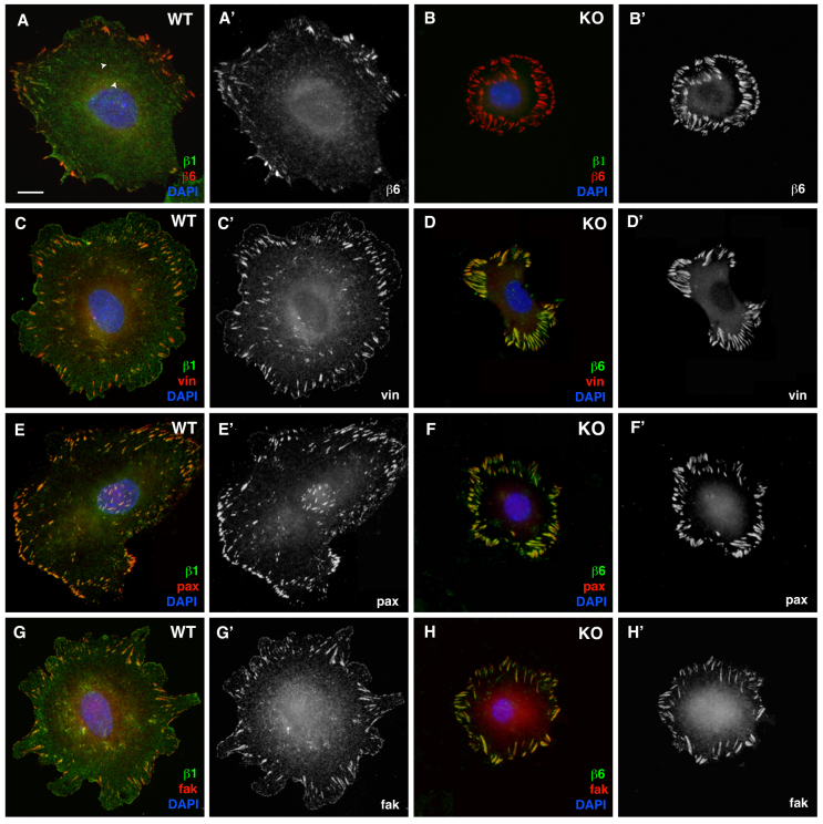
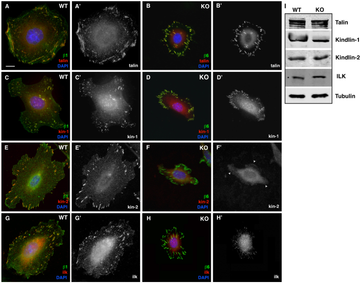
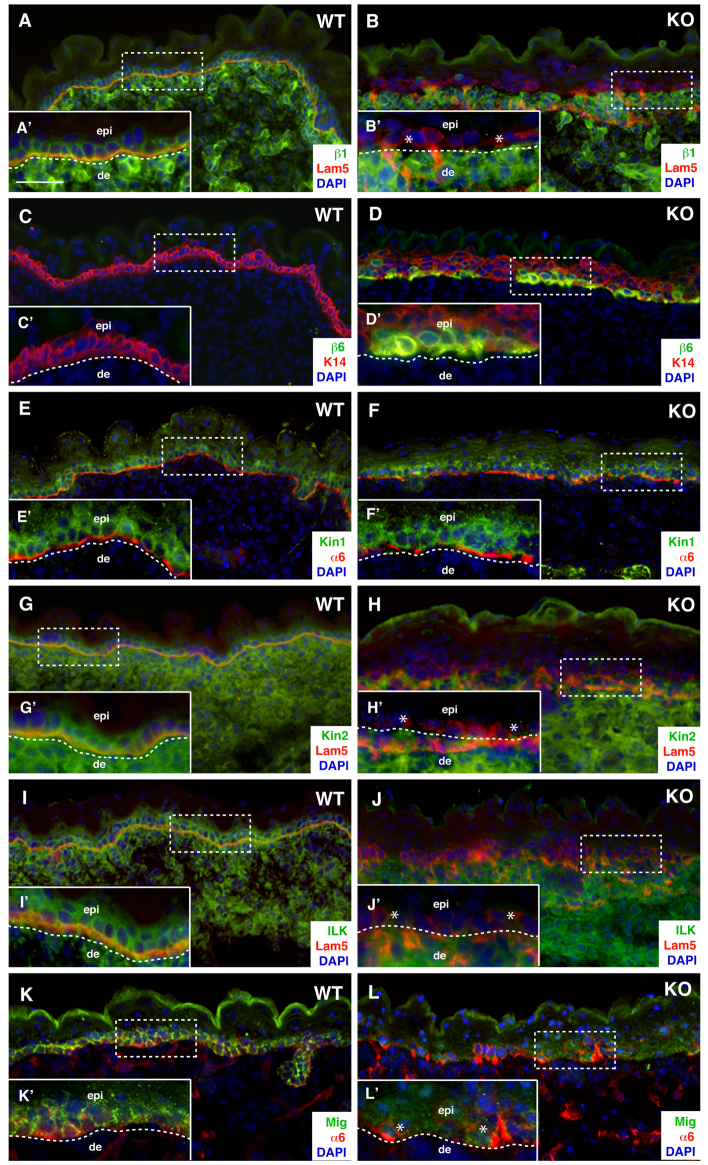
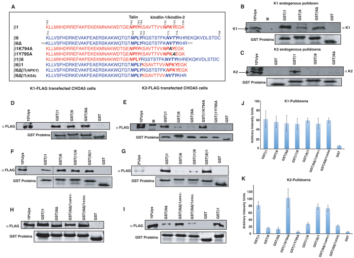
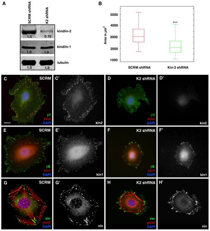
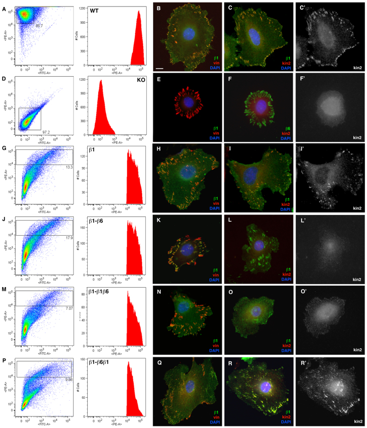
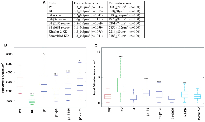
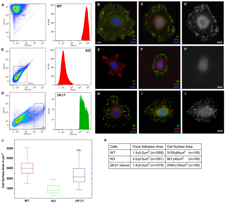
Integrin-β1-null keratinocytes can adhere to fibronectin through integrin αvβ6, but form large peripheral focal adhesions and exhibit defective cell spreading. Here we report that, in addition to the reduced avidity of αvβ6 integrin binding to fibronectin, the inability of integrin β6 to efficiently bind and recruit kindlin-2 to focal adhesions directly contributes to these phenotypes. Kindlins regulate integrins through direct interactions with the integrin-β cytoplasmic tail and keratinocytes express kindlin-1 and kindlin-2. Notably, although both kindlins localize to focal adhesions in wild-type cells, only kindlin-1 localizes to the integrin-β6-rich adhesions of integrin-β1-null cells. Rescue of these cells with wild-type and chimeric integrin constructs revealed a correlation between kindlin-2 recruitment and cell spreading. Furthermore, despite the presence of kindlin-1, knockdown of kindlin-2 in wild-type keratinocytes impaired cell spreading. Our data reveal unexpected functional consequences of differences in the association of two homologous kindlin isoforms with two closely related integrins, and suggest that despite their similarities, different kindlins are likely to have unique functions.


Figures

Fig. 1.
Fig. 1.
Localization of focal adhesion proteins in WT and KO cells. (AB′) WT (A,A′) and KO (B,B′) cells stained for β1 (green) and β6 (red), showing localization to the peripheral focal adhesions. The smaller focal complexes primarily contain β1 integrin (arrowheads, A). In all panels, the focal adhesions in the WT cells are stained with β1 (green), whereas those in the KO are stained with β6 (green) and co-stained in the WT cells with vinculin (C,C′) and), paxillin (E,E′ and FAK (G,G′) in red and in the KO cells with vinculin (D,D′) paxillin and FAK (H,H′) also in red. Scale bar: 10 μm.
Fig. 2.
Fig. 2.
Localization of β1-cytoplasmic-domain-interacting proteins in WT and KO cells. In all panels, WT focal adhesions are stained with β1 (green), whereas KO focal adhesions are stained with β6 (green) and co-stained in red with talin (A,B), kindlin-1 (C,D), kindlin-2 (E,F) and ILK (G,H). Talin and kindlin-1 strongly localize to the focal adhesions of both cell types (A′D′). Kindlin-2 fails to localize to the focal adhesions in the KO cell (arrowheads in F′) and there is very poor recruitment of ILK to the focal adhesion in the KO cells (H′). (I) Western blot analysis of WT and KO lysates with talin, kindlin-1, kindlin-2 and ILK. Tubulin was detected as a loading control. Scale bar: 10 μm.
Fig. 3.
Fig. 3.
Localization of β1-cytoplasmic-domain-interacting proteins in WT and KO skin. (A) WT and (B) KO skin sections stained with β1 (green) and laminin 5 (red). β1 is localized to the basal cells in WT (A′) and absent from the KO epidermis (B′). (C,C′) WT and (D,D′) KO skin sections stained with β6 (green) and K14 (red). There is de novo expression of β6 in the KO epidermis. (E) WT and (F) KO skin sections stained with kindlin-1 (green) and α6 integrin (red). Kindlin-1 is localized to the basal cells in WT (E′) and KO epidermis (F′). (G) WT and (H) KO skin sections stained with kindlin-2 (green) and laminin 5 (red). Kindlin-2 is not localized to the basal cells in the KO epidermis (asterisks in H′). (I) WT and (J) KO skin sections stained with ILK (green) and laminin 5 (red). ILK is not localized to the basal cells in the KO epidermis (asterisks in J′). (G) WT and (H) KO skin sections stained with migfilin (green) and α6 integrin (red). Migfilin is not localized to the basal cells in the KO epidermis (asterisks in L′). In all the WT and KO panels, the BM is demarcated with a dashed line at the dermal-–epidermal junction. The insets are higher magnification images of the dashed box. Scale bar: 200 μm (A–L); 100 μm (A′–L′). epi, epidermis; de, dermis.
Fig. 4.
Fig. 4.
Mapping the interaction between β-tails and kindlins using GST pull-down assays. (A) Sequences of the β1 and β6 cytoplasmic tails used to generate the GST constructs. Pull-down of endogenous kindlin-1 (B) and kindlin-2 (C) with GST–β1, GST–β6, GST–β6Δ and GST alone. Pull-down of FLAG-tagged kindlin-1 with GST–β1, GST–β6, GST–β6Δ and GST (D) and FLAG-tagged kindlin-2 with GST–β1, GST–β6, GST–β6Δ, GST–β1(K974A), GST–β1(Y795A) and GST (E) expressed in CHOA5 cells. Pull down of FLAG tagged kindlin-1 (F) and FLAG-tagged kindlin-2 (G) with GST–β1, GST–β6, GST–β1-β6, GST–β6-β1 and GST. (F). Pull down of FLAG tagged kindlin-1 (H) and FLAG-tagged kindlin-2 (I) with GST–β1, GST–β6Δ, GST–β6Δβ1(NPKY), GST–β6Δβ1(KSA) and GST. (F). Quantification of the amount of FLAG-tagged kindlin-1 (J) and kindlin-2 (K) pulled down by the various GST constructs. Results are normalized for loading against GST control (NIH ImageJ software). Data show means ± s.e. from three independent experiments.
Fig. 5.
Fig. 5.
Expression of kindlin-2 is important for cell spreading in keratinocytes. (A) Western blot analysis of lysates from cells expressing scrambled and kindlin-2 shRNA with kindlin-2 and kindlin-1. Tubulin was detected as a loading control. (B) Box plot of the distribution of the average cell area along the median (n=100) of scrambled and kindlin-2-knockdown cells (***P<0.001, compared with scrambled cells). Scrambled (C,C′) and kindlin-2-knockdown cells (D,D′) stained with β1 (green) and kindlin-2 (red). Scrambled (E,E′) and kindlin-2-knockdown cells (F,F′) stained with β6 (green) and kindlin-1 (red). Scrambled (G,G′) and kindlin-2-knockdown cells (H,H′) stained with vinculin (green) and phalloidin (red). Scale bar: 10 μm.
Fig. 6.
Fig. 6.
Rescue of β1-KO cell spreading correlates with targeting of kindlin-2 to focal adhesions. Surface expression of endogenous β1 on WT (A) and KO (D) cells assessed by FACS. (B,C) WT cells stained for β1, vinculin and kindlin-2, as indicated. (E,F) KO cells stained for β1, vinculin, β6 and kindlin-2, as indicated. (GR′) Surface expression of β1 on KO cells rescued with a full-length β1 (G), β1-β6 (J), β1–β1-β6 (M), or β1–β6-β1 (P) was assessed by FACS. Cells rescued with full-length β1 (H,I), β1-β6 (K,L), β1–β1-β6 (N,O), or β1–β6-β1 (Q,R) were stained for β1, vinculin and kindlin-2, as indicated. Scale bar: 10 μm.
Fig. 7.
Fig. 7.
Cell surface area and focal adhesion size are well correlated with the ability to recruit kindlin-2. (A) Table showing the mean focal adhesions and cell surface areas of the WT, KO, rescued cells, kindlin-2 KD and scrambled KD cells. (B) Box plot for average cell area (n=100) for WT, KO and rescued cells (***P<0.001, compared with WT cells, ^P>0.2, compared with WT). (C) Box plots for average focal adhesion area for WT, KO, rescued, kindlin-2 and scrambled KD cells (***P<0.001; FA significantly larger than WT).
Fig. 8.
Fig. 8.
Recruitment of Kindlin-2 by β6 integrin mediates cell spreading and focal complex formation. (A) Surface expression of endogenous β1 integrin on WT cells assessed by FACS. (B,C) WT cells stained for β1, vinculin and kindlin-2 (C′) as indicated. (D) Surface expression of endogenous β1 integrin on KO cells assessed by FACS. (E,F) KO cells stained with β1, vinculin, β6 and kindlin-2 (F′) as indicated. (G) Cells expressing high levels of β6-β1 were sorted based on high expression of GFP. Cells rescued with β6-β1 (H,I) were stained with β6 vinculin and kindlin-2 (I′) as indicated. (J) Box plot of the distribution of the average cell area along the median (n=100) of WT, KO and β6-β1-rescued cells (***P<0.001, compared with KO cells). (K) Table showing the mean focal adhesions and cell surface areas of the WT, KO and β1-β6-rescued cells. Scale bar: 10 μm.

References

    1. Ashton G. H., McLean W. H., South A. P., Oyama N., Smith F. J., Al-Suwaid R., Al-Ismaily A., Atherton D. J., Harwood C. A., Leigh I. M., et al. (2004). Recurrent mutations in kindlin-1, a novel keratinocyte focal contact protein, in the autosomal recessive skin fragility and photosensitivity disorder, Kindler syndrome. J. Invest. Dermatol. 122, 78-83 - PubMed
    1. Brakebusch C., Fässler R. (2005). beta 1 integrin function in vivo: adhesion, migration and more. Cancer Metastasis Rev. 24, 403-411 - PubMed
    1. Brakebusch C., Grose R., Quondamatteo F., Ramirez A., Jorcano J. L., Pirro A., Svensson M., Herken R., Sasaki T., Timpl R., et al. (2000). Skin and hair follicle integrity is crucially dependent on beta 1 integrin expression on keratinocytes. EMBO J. 19, 3990-4003 - PMC - PubMed
    1. Harburger D. S., Calderwood D. A. (2009). Integrin signalling at a glance. J. Cell Sci. 122, 159-163 - PMC - PubMed
    1. Harburger D. S., Bouaouina M., Calderwood D. A. (2009). Kindlin-1 and -2 directly bind the C-terminal region of beta integrin cytoplasmic tails and exert integrin-specific activation effects. J. Biol. Chem. 284, 11485-11497 - PMC - PubMed

Publication types

MeSH terms