. 2012 Apr;91(4):387-93.
doi: 10.1177/0022034512438401. Epub 2012 Feb 10.

Spry1 and spry2 are essential for development of the temporomandibular joint

Affiliations

Spry1 and spry2 are essential for development of the temporomandibular joint

P Purcell et al. J Dent Res. 2012 Apr.

Abstract

The temporomandibular joint (TMJ) is a specialized synovial joint essential for the function of the mammalian jaw. The main components of the TMJ are the mandibular condyle, the glenoid fossa of the temporal bone, and a fibrocartilagenous disc interposed between them. The genetic program for the development of the TMJ remains poorly understood. Here we show the crucial role of sprouty (Spry) genes in TMJ development. Sprouty genes encode intracellular inhibitors of receptor tyrosine kinase (RTK) signaling pathways, including those triggered by fibroblast growth factors (Fgfs). Using in situ hybridization, we show that Spry1 and Spry2 are highly expressed in muscles attached to the TMJ, including the lateral pterygoid and temporalis muscles. The combined inactivation of Spry1 and Spry2 results in overgrowth of these muscles, which disrupts normal development of the glenoid fossa. Remarkably, condyle and disc formation are not affected in these mutants, demonstrating that the glenoid fossa is not required for development of these structures. Our findings demonstrate the importance of regulated RTK signaling during TMJ development and suggest multiple skeletal origins for the fossa. Notably, our work provides the evidence that the TMJ condyle and disc develop independently of the mandibular fossa.


Conflict of interest statement

The authors declare that there is no conflict of interest.

Figures

Figure 1.
Figure 1.
Expression of members of the Fgf signaling pathway in the mouse TMJ region. Fgf signaling components were highly enriched in the mouse TMJ at E16.5 (Purcell et al., 2009). (A-I) Representative in situ hybridization in the mouse TMJ at E16.5. (A-D) Spry1, Spry2, and Spry4 are expressed in the lateral pterygoid and temporalis muscles, with Spry1 and Spry2 being highly expressed; Spry3 is not expressed. (E-H) Fgfr1 is expressed in the osteoblasts and periosteum of the condyle and fossa; Fgfr2 is expressed in perichondrium of the developing skeletal structures of the glenoid fossa and the condyle; Fgfr3 is present in the immature chondrocytes of the condyle; and Fgfr4 is expressed in the lateral pterygoid and temporalis muscles surrounding the TMJ. (I) Fgf6 is expressed in the lateral pterygoid and temporalis muscles. 10x magnification. c, condyle; f, glenoid fossa; lp, lateral pterygoid muscle; tm, temporalis muscle. Condyle and fossa have been outlined in black and muscles in red.
Figure 2.
Figure 2.
Spry1−/−;Spry2−/− embryos do not form a glenoid fossa. (A-H) Representative H&E staining from 4 different control and Spry1−/−;Spry2−/− (S1−/−;S2−/−) littermates at E14.5, E15.5, E16.5, and E18.5. (A,C,E,G) In control embryos, the glenoid fossa anlage (f*) and condyle (c) condensations are visible at E14.5, and the disc (d) is visible at E15.5 (black arrowhead). At all stages analyzed, the lateral pterygoid (lp) and temporalis muscles (tm) are detected (outlined in red). (B,D,F,H) In mutant embryos, the glenoid fossa is not visible at any stage, and its space is filled by an enlarged temporalis muscle. Instead of a fossa (f), a distal bony element forms at the region equivalent to the distal lateral tip of the fossa (i.e., zygomatic arch) in control TMJ (red arrowheads in A-D). The lateral pterygoid muscle is also enlarged starting at E15.5. The condyle and disc appear normal at all stages analyzed. (A-F) 10x magnification; (G,H) 4X magnification. (I,J) 3D reconstructions of microCT scans taken from 3 control and mutant heads at E18.5. To allow for better visibility of the glenoid fossa and the mandibular condyle, we removed the zygomatic bone and zygomatic processes of the temporal bone using the sculpting module in the Dolphin imaging software. MicroCT scans show a clear depression of the temporal bone in the control mice corresponding to the concave shape of the fossa (I; arrowhead). In mutant embryos, instead of the concave shape of the temporal bone, a flat surface is observed, confirming the absence of the glenoid fossa (J; arrowhead). The mandible is shown in yellow. The mandibular condylar process (c) of the mutant mouse is smaller by 50% in length and 25% in width compared with that in its control littermates. Analysis was restricted to embryos due to perinatal lethality of mutant mice. (K) The sizes of the lateral pterygoid and temporalis muscles at E18.5 were quantified. A 10-mm microscopic ruler (Klarmann Rulings, Inc.) was used to convert image pixel length to an SI unit of measurement (i.e., 1 mm = 533 pixels). Meckel’s cartilage (mc) showed no size difference between control and mutant mice. (L) Quantification of cell proliferation indicated by BrdU-positive cells at E17.5. n = 4; Student t test: *p < 0.05; **p < 0.01. c, condyle; d, disc; f, glenoid fossa; f*, fossa anlage; lp, lateral pterygoid muscle; mc, Meckel’s cartilage; tm, temporalis muscle.
Figure 3.
Figure 3.
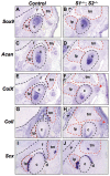
Condyle and disc develop normally in the absence of fossa. (A-J) In situ hybridization analysis on representative 10-µm serial coronal cryosections from 4 different control and Spry1−/−;Spry2−/− (S1−/−;S2−/−) littermates at E16.5. (A-D) Sox9 is localized to the proliferating chondrocytes, and Acan is localized to the cartilage of the condyle growth plate in control and mutant mice. (E,F) ColX is expressed in the zone of the hypertrophic chondrocytes of the condylar growth plate. (G,H) CoII expression is localized to the osteoblasts of the glenoid fossa and condyle in control and mutant mice. (I, J) Scx is expressed in tendons and tendon progenitors of the disc (black arrowheads) and muscle-bone contact sites. The lateral distal bony tip is indicated in mutant mice (F,H; red arrowheads). 10x magnification. c, condyle; f, glenoid fossa; f*, fossa anlage; lp, lateral pterygoid muscle; tm, temporalis muscle; Acan, aggrecan; ColX, collagen X; CoII, collagen I; Scx, scleraxis.
Figure 4.
Figure 4.
Model showing the requirement of sprouty genes in TMJ formation. Spry1, Spry2, Spry4, Fgfr4, and Fgf6 are expressed in the lateral pterygoid (lp) and temporalis (tm) muscle around the TMJ during embryonic development. Conditional inactivation of Spry1 and Spry2 in cartilage and bone indicate that these genes act cell-autonomously and exclusively in the muscles. We propose that SPRY1 and SPRY2 control muscle size through the regulation of signaling downstream of FGFR4. In Spry1−/−;Spry2−/− embryos, signaling downstream of FGFR4 is hyperactivated because of the lack of inhibition by sprouty proteins, resulting in uncontrolled muscle outgrowth. Since FGF6 is the key ligand for FGFR4 and is expressed by the muscle, it is likely that muscle growth is promoted by FGF6. The proposed mechanism in the temporalis muscle is also suggested to occur in the lateral pterygoid muscle. c, condyle; f, glenoid fossa; f*, fossa anlage; and z, zygomatic bone. Muscles are denoted in pink; condyle and fossa in blue; and disc in yellow. Light pink and light blue at E14.5 represent mesenchymal condensations for muscle and bone, respectively.

References

    1. Armand AS, Laziz I, Chanoine C. (2006). FGF6 in myogenesis. Biochim Biophys Acta 1763:773-778 - PubMed
    1. Avery JK. (2001). Oral development and histology. 3rd ed. New York, NY: Thieme Medical Publishers
    1. Basson MA, Akbulut S, Watson-Johnson J, Simon R, Carroll TJ, Shakya R, et al. (2005). Sprouty1 is a critical regulator of GDNF/RET-mediated kidney induction. Dev Cell 8:229-239 - PubMed
    1. Boros J, Newitt P, Wang Q, McAvoy JW, Lovicu FJ. (2006). Sef and Sprouty expression in the developing ocular lens: implications for regulating lens cell proliferation and differentiation. Semin Cell Dev Biol 17:741-752 - PMC - PubMed
    1. Brent AE, Tabin CJ. (2004). FGF acts directly on the somitic tendon progenitors through the Ets transcription factors Pea3 and Erm to regulate scleraxis expression. Development 131:3885-3896 - PubMed

Publication types

MeSH terms