

Preoperative localization of parathyroid lesion: diagnostic usefulness of color doppler ultrasonography
- PMID: 22328952
- PMCID: PMC3272690
Preoperative localization of parathyroid lesion: diagnostic usefulness of color doppler ultrasonography
Abstract
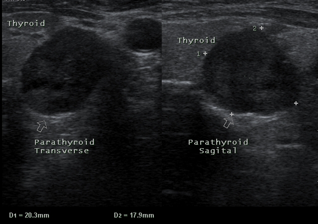
Introduction: Recently, minimally invasive parathyroidectomy (MIP) has been developed and is gaining popularity among surgeons. For this reason, preoperative localization is playing an important role to detect the precise location of the affected gland and to increase the success rate.
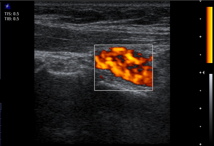
Material and methods: From June 2007 to June 2011, 56 consecutive patients (11 men and 45 women) with primary or secondary hyperparathyroidism in our center underwent Gray scale, color Doppler and 99m-Tc MIBI scan prior to operative management of parathyroid lesions.
Results: The sensitivity, specificity and accuracy of US and MIBI scan for pHPT was 88%, 94%, 91% and 70%, 100% and 85% respectively. In patients with sHPT, the sensitivity, specificity and accuracy of US and MIBI scan was 54%, 93%, 76% and 25%, 100% and 72.9% respectively. The overall sensitivity of combined US and MIBI scan in pHPT and sHPT was 97% and 45% respectively. The overall sensitivity, specificity and accuracy of CDUS in diagnosis of parathyroid lesions in pHPT and sHPT is 97%, 100%, 98.6% and 62%, 100% and 83% respectively.
Conclusion: The overall sensitivity and specificity of US and MIBI in preoperative localization of parathyroid adenoma in sHPT is lower than pHPT and performing CDUS can increases the overall sensitivity and specificity of imaging methods in accurate localization of parathyroid lesion.
Keywords: Preoperative localization; color doppler; diagnosis; parathyroid lesion; ultrasonography.
Figures
References
-
- Wermers RA, Khosla S, Atkinson EJ, Achenbach SJ, Oberg AL, Grant CS, Melton LJ., 3rd Incidence of primary hyperparathyroidism in Rochester, Minnesota, 1993-2001: an update on the changing epidemiology of the disease. J Bone Miner Res. 2006;21:171–177. - PubMed
-
- Mihai R, Wass JA, Sadler GP. Asymptomatic hyperparathyroidism-need for multicentre studies. Clinical Endocrinology. 2008;68:155–164. - PubMed
-
- Taniegra ED. Hyperparathyroidism. Am Fam Physician. 2004;69:333–339. - PubMed
-
- Marx SJ. Hyperparathyroid and hyperparathyroid disorders. N Engl J Med. 2000;343:1863–1875. - PubMed
LinkOut - more resources
Full Text Sources