. 2011 Dec;1(7):587-97.
doi: 10.1158/2159-8290.CD-11-0181.

Frequent alterations and epigenetic silencing of differentiation pathway genes in structurally rearranged liposarcomas

Affiliations

Frequent alterations and epigenetic silencing of differentiation pathway genes in structurally rearranged liposarcomas

Barry S Taylor et al. Cancer Discov. 2011 Dec.

Abstract

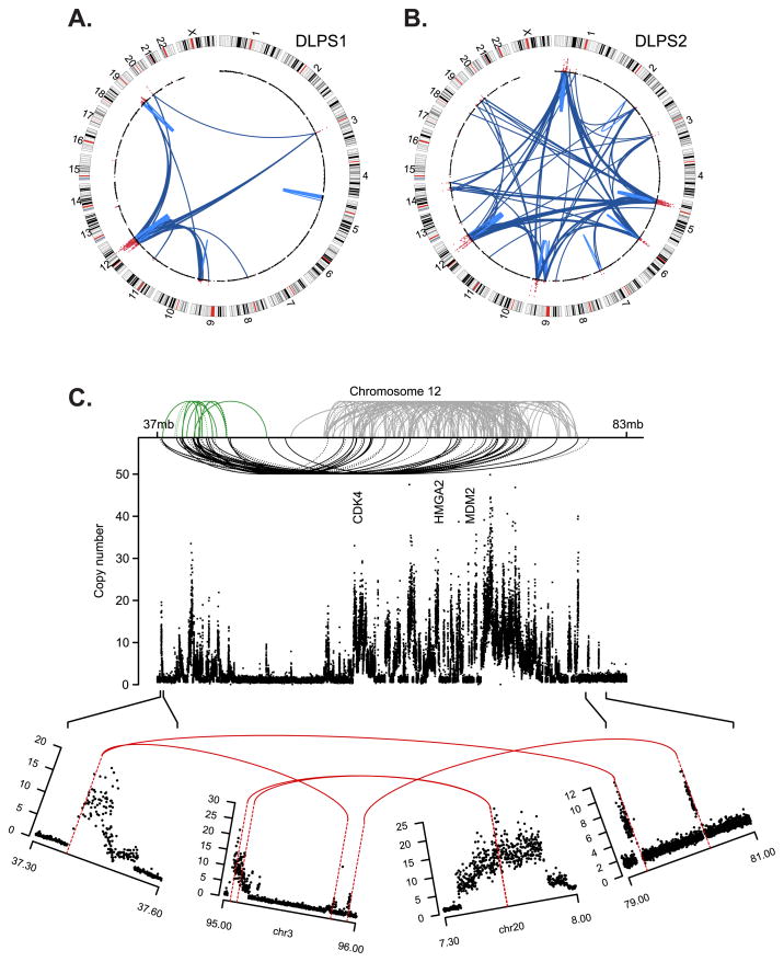
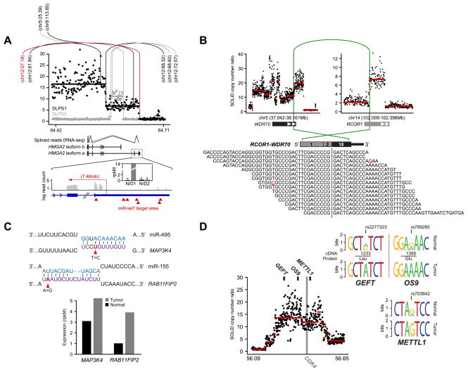
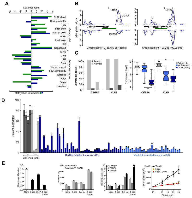
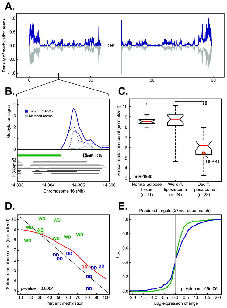
We explored diverse alterations contributing to liposarcomagenesis by sequencing the genome, exome, transcriptome, and cytosine methylome of a primary and recurrent dedifferentiated liposarcoma (DLPS) from distinct chemotherapy/radiotherapy-naïve patients. The liposarcoma genomes had complex structural rearrangements, but in different patterns, and with varied effects on the structure and expression of affected genes. While the point mutation rate was modest, integrative analyses and additional screening identified somatic mutations in HDAC1 in 8.3% of DLPS. Liposarcoma methylomes revealed alterations in differentiation pathway genes, including CEBPA methylation in 24% of DLPS. Treatment with demethylating agents, which restored CEBPA expression in DLPS cells, was anti-proliferative and pro-apoptotic in vitro and reduced tumor growth in vivo. Both genetic and epigenetic abnormalities established a role for small RNAs in liposarcomagenesis, typified by methylation-induced silencing of microRNA-193b in DLPS but not its well-differentiated counterpart. These findings reveal an unanticipated role for epigenetic abnormalities in DLPS tumors and suggest demethylating agents as potential therapeutics.

Significance: Multimodality sequence analysis of DLPS revealed recurrent mutations and epigenetic abnormalities critical to liposarcomagenesis and to the suppression of adipocyte differentiation. Pharmacologic inhibition of DNA methylation promoted apoptosis and differentiated DLPS cells in vitro and inhibited tumor growth in vivo, providing a rationale for investigating methylation inhibitors in this disease.

Keywords: DNA methylation; Dedifferentiated liposarcoma; adipocyte differentiation; histone deacetylase; microRNA.


Conflict of interest statement

Conflict of interest: No potential conflicts of interest to disclose.

Figures

Figure 1
Figure 1. Somatic rearrangements in two sarcomas
Structural rearrangements and DNA copy number alterations detected in two retroperitoneal liposarcoma genomes, a primary tumor, DLPS1 (A) and a local recurrence, DLPS2 (B). Chromosomes are plotted in the outer ring with the centromeres indicated in red. DNA copy number data inferred from whole-genome sequencing is indicated in the inner ring with genomic amplifications highlighted (red). Structural rearrangements are edges between two indicated loci, either intra-chromosomal (light blue) or inter-chromosomal (dark blue). C. The remodeling of chromosome 12 in DLPS1 is shown across ~46Mb of the q-arm. Three clusters (green, gray, and black) of both inverted and non-inverted intra-chromosomal rearrangements (dotted and solid, respectively) are shown spanning the progressive amplicon (copy number as indicated, y-axis). Three landmark genes (CDK4, HMGA2, and MDM2) are annotated. In the lower panel, the amplicon structure and rearrangement pattern of the 5’-most and 3’-most breakpoints are shown, indicating a circular structure including interspersed sequences from chromosomes 3 and 20, on top of which the complex pattern of intrachromosomal rearrangements (top) were likely acquired through successive rounds of unstable replication.
Figure 2
Figure 2. Diverse abnormalities manifest in RNA
A. The segmented copy number profile, inferred from whole-genome sequencing, of the HMGA2 locus (12q14.3) is plotted for the both DLPS1 (black) and DLPS2 (gray). Rearrangements are indicated (vertical/arc lines) and the position of their second breakpoint is annotated as megabases on the recipient chromosome. In red is the structural rearrangement in DLPS1 affecting the boundary between HMGA2 exon 5 and the 3’ UTR. Splice junction reads from RNA sequencing of DLPS1 (bottom panel) confirm that the long isoform (a) is expressed. A high-resolution view of the final exons and 3’ UTR of the transcript is shown; transcriptome read coverage confirms truncation and loss of the 3’ UTR from the rearrangement and significant over-expression of HMGA2 in DLPS1 (inset; gray and black are normal and tumor samples respectively). B. Rearrangement in DLPS2 pairing a simple intragenic amplicon in RCOR1 with a complex amplicon spanning WDR70. The RCOR1 intron 2 breakpoint juxtaposed the 5’ region including exon 2 with exon 18 and the amplified 3’ end of WDR70 (green arc). In total, 15 RNA sequencing reads supported the predicted fusion junction of this out-of-frame chimera. C. Somatic mutations in the 3’ UTRs of MAP3K4 and RAB11FIP2 in DLPS2 that were detected from RNA but not exome sequencing fall in the seed regions of conserved target sites complementary to miR-495 and miR-155 respectively (purple, microRNA sequences are shown above in blue). Both genes have elevated expression in DLPS2 compared to their matched normal tissue (bottom, measured as rpkM), consonant with release from microRNA repression. D. Allele-specific expression of GEFT, OS9, and METTL1, three genes adjacent to the CDK4 oncogene on 12q13.3–14.1, was detected from heterozygous exonic variants from RNA sequencing in DLPS1 (reflected as sequence logos inferred from spanning reads) and is attributable to a single complex genomic amplification.
Figure 3
Figure 3. Core promoter methylation in mediators of adipogenesis
A. Patterns of somatic methylation enriched or depleted (measured by log odds ratio relative to background sequence in the human genome) among 19 sequence contexts including the canonical gene cassette. Tumor-specific increases and decreases of methylation are green and blue, respectively. B. Methylation of CEBPA and KLF4 is increased in tumors (blue), but not their matched normal adipose tissues (gray) in upstream regions or in regions spanning adjacent CpG islands (green). Methylation signal for each tumor is plotted for both the positive and negative strands (dotted and dashed respectively) and combined into total signal (solid) for each sample. Also indicated is the distance between the peak of methylation in both promoters and their respective transcription starts sites. C. CEBPA and KLF4 expression, inferred from RNA sequencing in the methylated samples (normalized rpkM) indicates their reduction in DLPS1 and DLPS2 compared to their matched normal adipose tissues (left panel). On the right, expression measured by microarray in a cohort of 115 tissues (as shown; starred, p-values < 10−12, ANOVA). D. CEBPA promoter methylation status (average percent methylated, two biological replicates assessed by bisulfite pyrosequencing, error bars represent standard deviation) in 8 cell lines (dark and light gray are dedifferentiated and well-differentiated cell lines, respectively) and 72 tumors indicated that CEBPA methylation is high in cell lines and a subset of DLPS, but absent from WLPS tumors. E. Proliferation of DDLS8817 dedifferentiated liposarcoma cells after treatment with 5-aza, SAHA, or the combination of both (left; mean ± propagated error). Proliferation levels are shown relative to untreated cells. The percentage of apoptotic cells is shown (middle, left; measured by annexin V and 7-AAD staining, mean percent positive ± standard deviation) in untreated and drug-treated DDLPS8817 cells. Expression of early, intermediate, and late markers of differentiation (perilipin, FABP4, and adipsin respectively) were measured by RT-PCR (mean ± standard deviation) in the presence of drug and shown relative to the level in untreated cells (middle, right). The growth of DLPS tumors in mice (DDLS8817 xenografts) treated with 5-aza (decitabine), 5-aza plus SAHA, or vehicle (right; mean tumor volume ± standard error of the mean, n = 5 mice/group) was also analyzed.
Figure 4
Figure 4. Epigenetic regulation of miR-193b in liposarcomagenesis
A. The density of methylation on chromosome 16 in DLPS1 (blue) and its matched normal adipose tissue (gray). B. Methylation in the primary tumor (solid blue, positive and negative strands are dotted and dashed respectively) and matched normal adipose tissue (gray) in the region of the putative promoter [the position of enrichment of histone H3K4me3 in nine human cell types (20) is indicated by horizontal lines] of miR-193b overlapping the shore of a CpG island (green bar). C. Expression of miR-193b in normal adipose tissue samples, WLPS, and DLPS tumors determined by small RNA sequencing. The tumor in which methylation was observed (panel B) is highlighted (starred; p-value < 10−9, Student’s t-test). D. Expression of miR-193b as a function of percent methylation in the putative miR-193b promoter in a panel of both well-differentiated (WD, green) and dedifferentiated (DD, blue; DLPS1 is in red ) liposarcomas [n = 17; the diagonal is indicated by dotted line; the red line is the regression (loess)]. E. Cumulative distributions of expression changes of predicted miR-193b target genes (≥ 7 bp seed match length, n = 547 genes with expression data) between DLPS tumors and normal adipose tissues (green) or WLPS tumors and normal adipose tissues (blue) indicate that a greater number of predicted targets had increased expression in DLPS tumors with methylated miR-193b than expected by chance (p-value as indicated, Kolmogorov-Smirnov).

Comment in

References

    1. Fletcher C, Unni K, Mertens F. Pathology and genetics of tumors of soft tissue and bone. International Agency for Research on Cancer Press; Lyon: 2002.
    1. Pedeutour F, Forus A, Coindre JM, Berner JM, Nicolo G, Michiels JF, et al. Structure of the supernumerary ring and giant rod chromosomes in adipose tissue tumors. Genes Chromosomes Cancer. 1999;24:30–41. - PubMed
    1. Barretina J, Taylor BS, Banerji S, Ramos AH, Lagos-Quintana M, Decarolis PL, et al. Subtype-specific genomic alterations define new targets for soft-tissue sarcoma therapy. Nat Genet. 2010;42:715–21. - PMC - PubMed
    1. Campbell PJ, Yachida S, Mudie LJ, Stephens PJ, Pleasance ED, Stebbings LA, et al. The patterns and dynamics of genomic instability in metastatic pancreatic cancer. Nature. 2010;467:1109–13. - PMC - PubMed
    1. Stephens PJ, McBride DJ, Lin ML, Varela I, Pleasance ED, Simpson JT, et al. Complex landscapes of somatic rearrangement in human breast cancer genomes. Nature. 2009;462:1005–10. - PMC - PubMed

Publication types