. 2012 Dec 18;45(12):2114-24.
doi: 10.1021/ar200294y. Epub 2012 Feb 14.

Aqueous phase separation as a possible route to compartmentalization of biological molecules

Affiliations
Free PMC article

Aqueous phase separation as a possible route to compartmentalization of biological molecules

Christine D Keating. Acc Chem Res. .
Free PMC article

Abstract

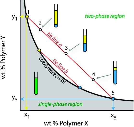
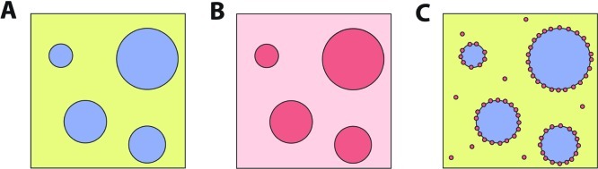
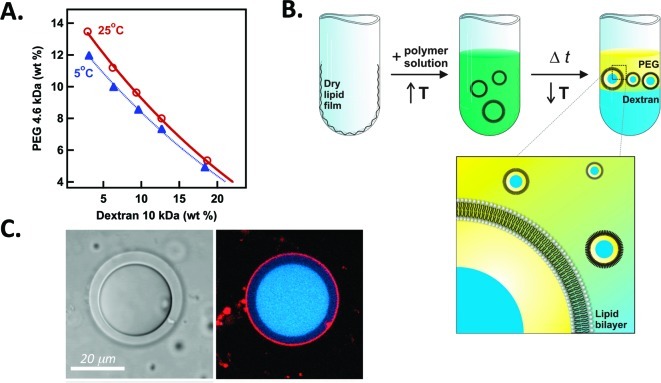
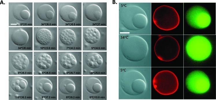
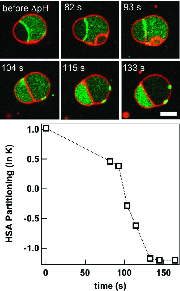
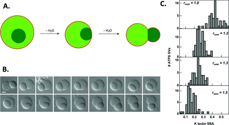
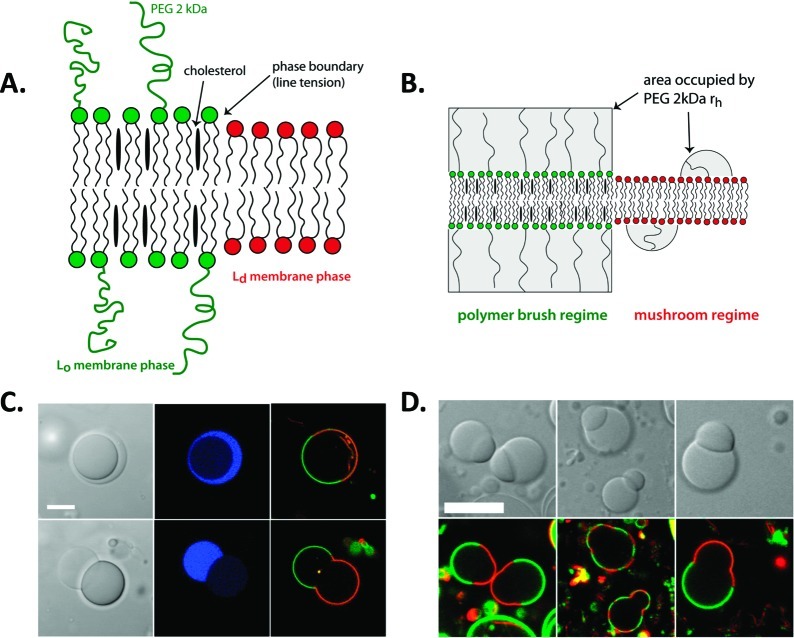
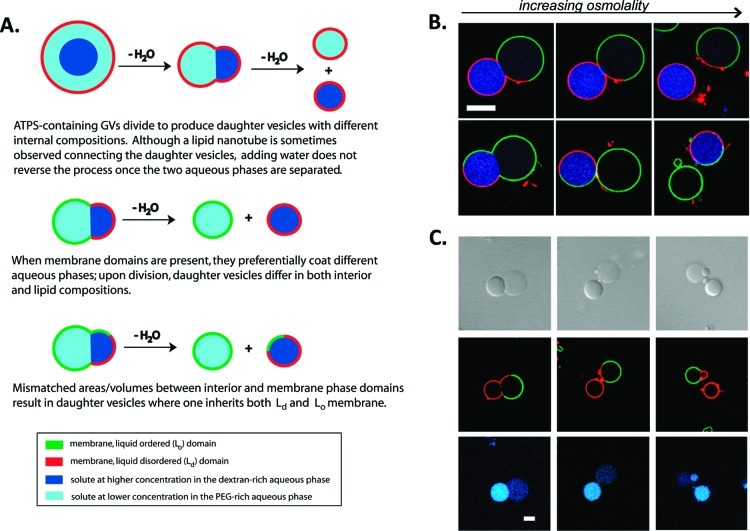
How could the incredible complexity of modern cells evolve from something simple enough to have appeared in a primordial soup? This enduring question has sparked the interest of researchers since Darwin first considered his theory of natural selection. Organic molecules, even potentially functional molecules including peptides and nucleotides, can be produced abiotically. Amphiphiles such as surfactants and lipids display remarkable self-assembly processes including the spontaneous formation of vesicles resembling the membranes of living cells. Nonetheless, numerous questions remain. Given the presumably dilute concentrations of macromolecules in the prebiotic pools where the earliest cells are thought to have appeared, how could the necessary components become concentrated and encapsulated within a semipermeable membrane? What would drive the further structural complexity that is a hallmark of modern living systems? The interior of modern cells is subdivided into microcompartments such as the nucleoid of bacteria or the organelles of eukaryotic cells. Even within what at first appears to be a single compartment, for example, the cytoplasm or nucleus, chemical composition is often nonuniform, containing gradients, macromolecular assemblies, and/or liquid droplets. What might the internal structure of intermediate evolutionary forms have looked like? The nonideal aqueous solution chemistry of macromolecules offers an attractive possible answer to these questions. Aqueous polymer solutions will form multiple coexisting thermodynamic phases under a variety of readily accessible conditions. In this Account, we describe aqueous phase separation as a model system for biological compartmentalization in both early and modern cells, with an emphasis on systems that have been encapsulated within a lipid bilayer. We begin with an introduction to aqueous phase separation and discuss how this phenomenon can lead to microcompartmentalization and could facilitate biopolymer encapsulation by partitioning of solutes between the phases. We then describe primitive model cells based on phase separation inside lipid vesicles, which mimic several basic properties of biological cells: microcompartmentation, protein relocalization in response to stimulus, loss of symmetry, and asymmetric vesicle division. We observe these seemingly complex phenomena in the absence of genetic molecules, enzymes, or cellular machinery, and as a result these processes could provide clues to possible intermediates in the early evolution of cell-like assemblies.


Figures

Figure 1
Figure 1
Generic phase diagram for an aqueous solution of two neutral polymers. The concentration of each polymer in the top and bottom phase is given by the intersection of the tie line on which that composition lies with the coexistence curve. Here, points 2, 3, and 4 are above the coexistence curve and therefore exist as two phases. These points lie on the same tie line and consequently their top and bottom phases are each given by points 1 (top phase) and 5 (bottom phase) but differ in volume as indicated in the illustration.
Figure 2
Figure 2
Agitation of a bulk ATPS results in micrometer-scale droplets of one phase suspended in the other phase (A). Two examples of partitioning are shown: partitioning between the two aqueous phases with no significant accumulation at the interface (B) and particulate partitioning between one of the phases and the interface (C).
Figure 3
Figure 3
Preparation of artificial cells in which ATPS are encapsulated by a lipid membrane. (A) phase diagram for a PEG/dextran ATPS showing temperature dependence. (B) Vesicles are formed by gentle hydration in a warm polymer solution, after which cooling leads to phase separation. Reprinted with permission from ref (33). Copyright 2005 National Academy of Sciences. (C) Partitioning in an ATPS-containing giant vesicle. Fluorescently labeled dextran is shown in blue, and lipid in red. Image acquired by M. Andes-Koback.
Figure 4
Figure 4
Reversible microcompartmentalization in an ATPS-containing vesicle. (A) Optical microscopy images during heating and subsequent cooling show phase transitions in the interior ATPS. (B) Fluorescence microscopy indicates the location of the lipid membrane (red) and a protein concanavalin A (green) before and after temperature changes. Scale bars are 10 μm. Reprinted with permission from ref (33). Copyright 2005 National Academy of Sciences.
Figure 5
Figure 5
Protein migration between aqueous phase microcompartments within a giant lipid vesicle. Human serum albumin (HSA, green) moves from the PEG-rich compartment and ATPS interface at pH 4.1 to the dextran-rich compartment at pH 6.5. Lipid membrane is shown in red; scale bar is 5 μm. Reprinted with permission from ref (28). Copyright 2010 American Chemical Society.
Figure 6
Figure 6
Response of ATPS-containing GVs to osmotic stress. (A) Schematic illustration of budding process. (B) The budding transition is reversible. Here, water is added to the external medium of a budded vesicle, causing a volume increase and retraction of the dextran-rich bud. Subsequent exposure to hypertonic sucrose solution regenerates the budded structure. Scale bar is 10 μm. (C) Histograms for partitioning of a protein, soybean agglutinin as a function of external osmolality; final external/initial internal osmolality ratios, rosm, are given for each panel. Reprinted with permission from ref (38). Copyright 2008 American Chemical Society.
Figure 7
Figure 7
Correspondence between interior aqueous microcompartments and membrane phase domains. Illustrations of (A) phase separation in ternary lipid mixture, and (B) higher surface density of PEGylated headgroups leading to polymer brush regime for Lo domain while surface densities for Ld domain are in the mushroom regime. (C) Optical microscope images for an ATPS-containing GV before and after undergoing a budding transition. (D) Effect of temperature on lipid phase domain distribution on budded GVs. Blue indicates the PEG-rich aqueous phase, green fluorescence indicates PEGylated Lo lipid domain, and red indicates Ld lipid domain. Reprinted with permission from ref (42). Copyright 2008 American Chemical Society.
Figure 8
Figure 8
Asymmetric fission of ATPS-containing GV. (A) Illustration of outcomes based on initial conditions. (B) Fluorescence microscopy showing two division events yielding nonidentical daughter vesicles, one of which is already polarized due to the presence of both membrane domains. (C) Budding of a daughter vesicle after initial fission event. Panels top to bottom are transmitted light (DIC), membrane fluorescence, and interior protein fluorescence. Red indicates Ld domain lipid (DOPE-rhodamine), and green indicates protein bound to Lo domain lipid (streptavidin-Alexa 488, bound to DSPE-PEG 2000-biotin), and blue indicates a protein, SBA-Alexa 647, which is partitioned into the dextran-rich interior aqueous phase. T = 5 °C. Scale bar is 10 μm. Reprinted with permission from ref (44). Copyright 2011 American Chemical Society.

References

    1. Follmann H.; Brownson C. Darwin’s warm little pond revisited: from molecules to the origin of life. Naturwissenschaften 2009, 96, 1265–1292. - PubMed
    1. Pohorille A.; Deamer D. Self-assembly and function of primitive cell membranes. Res. Microbiol. 2009, 160, 449–456. - PubMed
    1. Noireaux V.; Libchaber A. A vesicle bioreactor as a step toward an artificial cell assembly. Proc. Natl. Acad. Sci. U.S.A. 2004, 101, 17669–17674. - PMC - PubMed
    1. Kurihara K.; Tamura M.; Shohda K.-i.; Toyota T.; Suzuki K.; Sugawara T. Self-reproduction of supramolecular giant vesicles combined with the amplification of encapsulated DNA. Nat. Chem 2011, 3, 775–781. - PubMed
    1. The Origins of Life. Cold Spring Harbor Perspectives in Biology; Deamer D., Szostak J. W., Eds.; Cold Spring Harbor Press: Cold Spring Harbor, NY, 2010; 318 pp.

Publication types