. 2012 Jan 30;20(3):2706-16.
doi: 10.1364/OE.20.002706.

Time-resolved protein nanocrystallography using an X-ray free-electron laser

Affiliations

Time-resolved protein nanocrystallography using an X-ray free-electron laser

Andrew Aquila et al. Opt Express. .

Abstract

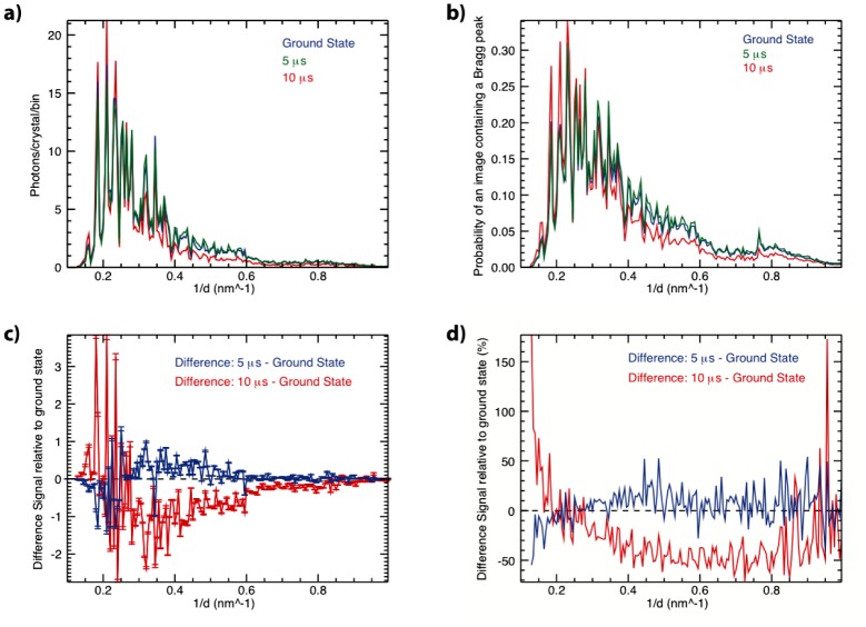
We demonstrate the use of an X-ray free electron laser synchronized with an optical pump laser to obtain X-ray diffraction snapshots from the photoactivated states of large membrane protein complexes in the form of nanocrystals flowing in a liquid jet. Light-induced changes of Photosystem I-Ferredoxin co-crystals were observed at time delays of 5 to 10 µs after excitation. The result correlates with the microsecond kinetics of electron transfer from Photosystem I to ferredoxin. The undocking process that follows the electron transfer leads to large rearrangements in the crystals that will terminally lead to the disintegration of the crystals. We describe the experimental setup and obtain the first time-resolved femtosecond serial X-ray crystallography results from an irreversible photo-chemical reaction at the Linac Coherent Light Source. This technique opens the door to time-resolved structural studies of reaction dynamics in biological systems.


Figures

Fig. 1
Fig. 1
Experimental setup and intersection of the 3 interacting beams: X-rays, pump laser, and the liquid jet containing the co-crystals. The upper left insert shows the thermal glow of the 2 keV X-rays interacting with the liquid jet. The lower insert shows the scatter from the frequency-doubled Nd:YAG pump laser from the liquid jet. The overlap of the two beams (liquid jet and X-rays) can be seen, with the pump laser intersecting the liquid jet of co-crystals and extending upstream towards the nozzle. The pump laser is extended upstream of the X-ray pulse to compensate for the approximately 130 μm travel of the crystals between the pump and probe pulses for the 10 μs delay time.
Fig. 2
Fig. 2
Single-shot, single crystal diffraction patterns, from the pnCCD detectors, of PSI-fd (top) in the dark state, (middle) in the 5 μs pumped state, and (bottom) the 10 μs pumped state. The images on the right side are the same as the corresponding left side image, except predicted peak locations from indexing are shown in gray circles. Partial reflections were observed containing between 10 to 500 photons per peak with a background level of around 2 photons caused by water scatter, fluorescence, and detector noise.
Fig. 3
Fig. 3
(a) The 1D virtual powder patterns for the ground state and two positive time delays for PSI-fd co-crystals. The intensities were linearly scaled to minimize the signal difference between for the different excited delays to the ground state. The powder patterns show the average number of photons scattered per crystal hit with in a histogram bin size of q = 0.005 nm−1. The area under the curve corresponds to 393.3, 406.6, and 378.1 average diffracted photons per crystal detected for the dark, 5 μs, and 10 μs data sets respectively. (b) Counting statistics of the number of peaks used in the virtual powder pattern recorded as a function of 1/d. The area under the curve corresponds to the average number of peaks per crystal diffraction pattern of 10.8, 11.5, and 9.9 peaks/pattern for the dark, 5 μs, and 10 μs data sets respectively. (c) Relative difference signal between the ground state and the excited states as a function of resolution. Error bars on the data points are 3σ errors. (d) Difference signal expressed as a percentage change.

References

    1. Schotte F., Soman J., Olson J. S., Wulff M., Anfinrud P. A., “Picosecond time-resolved X-ray crystallography: probing protein function in real time,” J. Struct. Biol. 147(3), 235–246 (2004).10.1016/j.jsb.2004.06.009 - DOI - PubMed
    1. Wöhri A. B., Katona G., Johansson L. C., Fritz E., Malmerberg E., Andersson M., Vincent J., Eklund M., Cammarata M., Wulff M., Davidsson J., Groenhof G., Neutze R., “Light-induced structural changes in a photosynthetic reaction center caught by Laue diffraction,” Science 328(5978), 630–633 (2010).10.1126/science.1186159 - DOI - PubMed
    1. Graber T., Anderson S., Brewer H., Chen Y. S., Cho H. S., Dashdorj N., Henning R. W., Kosheleva I., Macha G., Meron M., Pahl R., Ren Z., Ruan S., Schotte F., Srajer V., Viccaro P. J., Westferro F., Anfinrud P., Moffat K., “BioCARS: a synchrotron resource for time-resolved X-ray science,” J. Synchrotron Radiat. 18(4), 658–670 (2011).10.1107/S0909049511009423 - DOI - PMC - PubMed
    1. Moffat K., “The frontiers of time-resolved macromolecular crystallography: movies and chirped X-ray pulses,” Faraday Discuss. 122, 65–77, discussion 79–88 (2003).10.1039/b201620f - DOI - PubMed
    1. Moffat K., Szebenyi D., Bilderback D., “X-ray Laue Diffraction from Protein Crystals,” Science 223(4643), 1423–1425 (1984).10.1126/science.223.4643.1423 - DOI - PubMed

Publication types