

Review
doi: 10.1097/WNO.0b013e3182474548.
Shedding light on photophobia
Affiliations
- PMID: 22330853
- PMCID: PMC3485070
- DOI: 10.1097/WNO.0b013e3182474548
Item in Clipboard
Review
Shedding light on photophobia
J Neuroophthalmol.
2012 Mar.
Abstract
Photophobia is a common yet debilitating symptom seen in many ophthalmic and neurologic disorders. Despite its prevalence, it is poorly understood and difficult to treat. However, the past few years have seen significant advances in our understanding of this symptom. We review the clinical characteristics and disorders associated with photophobia, discuss the anatomy and physiology of this phenomenon, and conclude with a practical approach to diagnosis and treatment.
Conflict of interest statement
Conflict of interest statement: The authors report no conflicts of interest.
Figures
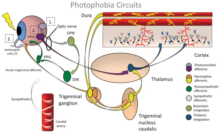
1. Ganglion cells project light-related signaling to the olivary pretectal nucleus (OPN; light green). OPN projections activate superior salivatory nucleus (SSN; dark green), which via pterygopalatine ganglion, causes ocular vasodilation and activation of ocular trigeminal afferents (orange) which are heavily expressed on blood vessels. These afferents, with cell bodies in the trigeminal ganglion, project to trigeminal nucleus caudalis, thalamus and cortex. 2. Intrinsically photosensitive retinal ganglion cells (IPRGCs) project directly to thalamic neurons (blue) that also receive intracranial nociceptive afferent signal (yellow neurons in trigeminal ganglion and trigeminal nucleus caudalis. Thalamic neurons fire in response to light and pain stimuli. Their output projects diffusely to sensory and association cortex. 3. Melanopsin-containing, intrinsically photosensitive ganglion-like cells have been identified in rodent iris. These afferents may explain the fact that light can activate trigeminal blink reflex even after the optic nerve (through which circuits 1. and 2. pass) has been sectioned. Note that all three circuits may interact at different locations. (created from references –86, 91).
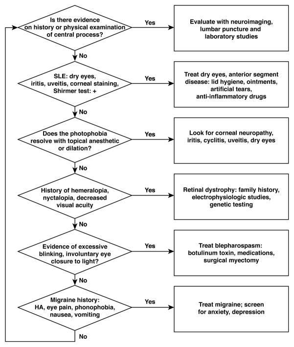
An approach to the patient with photophobia.
Comment in
-
Relief of refractory photo-oculodynia with botulinum toxin.J Neuroophthalmol. 2012 Sep;32(3):293. doi: 10.1097/WNO.0b013e3182585b5d. J Neuroophthalmol. 2012. PMID: 22549562 No abstract available.
References
-
- Classification and diagnostic criteria for headache disorders, cranial neuralgias and facial pain. Headache Classification Committee of the International Headache Society. Cephalalgia. 1988;8 (Suppl 7):1–96. - PubMed
-
- Headache Classification Committee of the International Headache Society. The International Classification of Headache Disorders. Cephalalgia. 2004;24:1–160. - PubMed
-
- Trobe JD. Photophobia in anterior visual pathway disease. J Neuroophthalmol. 2002;22:1–2. - PubMed
-
- Webster’s New World Dictionary. Cleveland: The World Publishing Company; 1968.
-
- Lebensohn J. The nature of photophobia. Arch Ophthalmol. 1934;12:380–383.
Publication types
MeSH terms
Grants and funding
LinkOut - more resources
Full Text Sources
Other Literature Sources
Medical
Miscellaneous
