. 2012 Jan;51(1):50-7.

Sedation or inhalant anesthesia before euthanasia with CO2 does not reduce behavioral or physiologic signs of pain and stress in mice

Affiliations

Sedation or inhalant anesthesia before euthanasia with CO2 does not reduce behavioral or physiologic signs of pain and stress in mice

Helen Valentine et al. J Am Assoc Lab Anim Sci. 2012 Jan.

Abstract

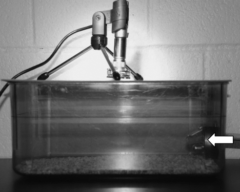
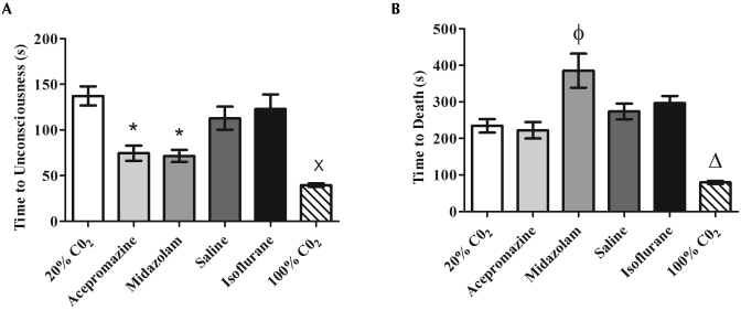
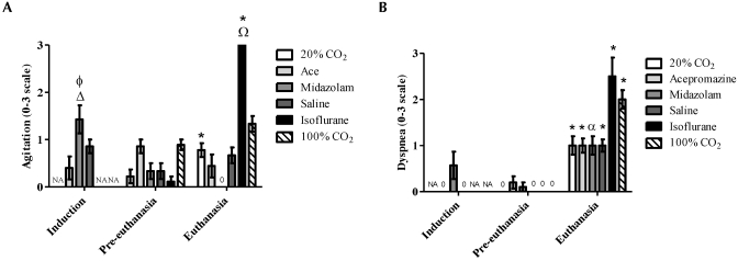
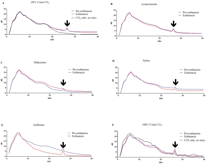
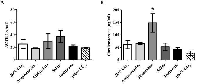
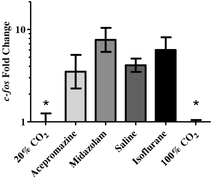
CO(2) administration is a common euthanasia method for research mice, yet questions remain regarding whether CO(2) euthanasia is associated with pain and stress. Here we assessed whether premedication with acepromazine, midazolam, or anesthetic induction with isoflurane altered behavioral and physiologic parameters that may reflect pain or stress during CO(2) euthanasia. Mice were assigned to 1 of 6 euthanasia groups: CO(2) only at a flow rate of 1.2 L/min which displaces 20% of the cage volume per minute (V/min; control group); premedication with acepromazine (5 mg/kg), midazolam (5 mg/kg), or saline followed by 20% V/min CO(2); induction with 5% isoflurane followed by greater than 100% V/min CO(2) (>6L/min); and 100% V/min CO(2) only (6 L/min). Measures included ultrasonic sound recordings, behavioral analysis of video record- ings, plasma ACTH and corticosterone levels immediately after euthanasia, and quantification of c-fos from brain tissue. Compared with 20% V/min CO(2) alone, premedication with acepromazine or midazolam did not significantly alter behavior but did induce significantly higher c-fos expression in the brain. Furthermore, the use of isoflurane induction prior to CO(2) euthanasia significantly increased both behavioral and neuromolecular signs of stress. The data indicate that compared with other modalities, 20% V/min CO(2) alone resulted in the least evidence of stress in mice and therefore was the most humane euthanasia method identified in the current study.


Figures

Figure 1.
Figure 1.
The euthanasia set-up. Mice remained in the home cage, CO2 was administered through the lixit port (arrow), and an ultrasonic microphone was placed over the cage.
Figure 2.
Figure 2.
Times (mean ± SEM) to (A) unconsciousness and (B) death were analyzed for each euthanasia group. Mice given either acepromazine or midazolam or euthanized with 100% V/min CO2 experienced the most rapid time to unconsciousness, but only mice euthanized with 100% V/min CO2 experienced a significantly more rapid death compared with other groups. Despite a shorter time to unconsciousness, mice euthanized with midazolam experienced a significantly increased time to death compared with that of mice that received 20% V/min CO2. (A) *, Significant (P < 0.05) decrease in time compared with values for 20% V/min CO2 and isoflurane groups; ×, significant (P < 0.05) decrease in time compared with 20% V/min CO2, saline, and isoflurane groups; (B) Φ, significant (P < 0.05) increase in time compared with values for 20% V/min CO2, acepromazine, and saline groups; Δ, significant (P < 0.05) decrease in time compared with values for all other groups.
Figure 3.
Figure 3.
A blinded observer examined videotapes taken at induction, preeuthanasia, and euthanasia and scored levels of (A) agitation (increased and altered activity) and (B) dyspnea (increased respiratory effort) on a scale of 0 to 3 (0, none; 1, mild; 2, moderate; 3, severe). For both agitation and dyspnea, mice anesthetized with isoflurane displayed the highest mean scores at euthanasia. Regardless of treatment, all groups displayed significant (P < 0.05) dyspnea at the time of euthanasia compared with either preeuthanasia or induction levels. NA, no applicable data. (A) Δ, Significant (P < 0.05) increase in agitation compared with values at preeuthanasia and euthanasia time points; Φ, significant (P < 0.05) increase in agitation compared with that of with mice induced with acepromazine; *, significant (P < 0.05) increase in agitation compared with that of respective preeuthanasia score; Ω, significant (P < 0.05) increase in agitation at euthanasia compared with that of mice treated with 20% V/min CO2, acepromazine, midazolam, or saline. (B) *, Significant (P < 0.05) increase in dyspnea compared with that of respective preeuthanasia and induction scores (if applicable); α, indicates a significant increase in dyspnea compared with that for the respective preeuthanasia score.
Figure 4.
Figure 4.
Spectrograms of ultrasonic sound recordings for each euthanasia group were graphed. Preeuthanasia recordings were generated for 2 min prior to administration of gas. Euthanasia recordings were initiated at the beginning of gas administration until all mice in the cage were unconscious. Each figure represents the averaged values from 2 cages of mice from each group. All groups displayed a peak at 26.5 kHz (arrow), during both recordings, which was absent when mice were not in the cage. This peak represents the only peak readily attributable to vocalization. Mice (A) euthanized with 20% V/min CO2 or (B) administered acepromazine had identical baseline preeuthanasia and euthanasia recordings. In contrast, (C) mice treated with midazolam had lower dB recordings in the 10- to 25-kHz range during euthanasia, consistent with heavy sedation and decreased movement, but not a decrease at the 26.5-kHz vocalization peak. Mice given (D) saline, (E) isoflurane, or (F) 100% V/min CO2 had higher amplitude recordings at the vocalization peak, consistent with increased vocalization at euthanasia. (E) Isoflurane euthanasia also produced a large degree of background noise (10- to 26.5-kHz range), consistent with increased movement and agitation noted in this group.
Figure 5.
Figure 5.
Plasma levels (mean ± SEM) of (A) ACTH and (B) corticosterone for each euthanasia group were evaluated. The mice that received midazolam displayed a significant (*, P < 0.05) increase in corticosterone but not ACTH; this result was suggestive of a hypoxic ACTH-independent activation of adrenal steroidogenesis. This finding was consistent with the increased time to death following unconsciousness in this group.
Figure 6.
Figure 6.
Relative mean expression (mean ± SEM) of c-fos in the brains of mice compared with the 20% V/min CO2 control group calculated by using the ΔΔct method. Both the 20%- and 100%-CO2–treated groups expressed significantly (*, P < 0.05) less c-fos than did all other groups.

Comment in

References

    1. American Veterinary Medical Association. [Internet]. 2007. AVMA guidelines on euthanasia, 2007 update. [Cited 16 June 2011]. Available at: http://www.avma.org/issues/animal_welfare/euthanasia.pdf.
    1. Anton F, Euchner I, Handwerker HO. 1992. Psychophysical examination of pain induced by defined CO2 pulses applied to the nasal mucosa. Pain 49:53–60 - PubMed
    1. Artwohl J, Brown P, Corning B, Stein S. 2006. Report of the ACLAM Task Force on Rodent Euthanasia. J Am Assoc Lab Anim Sci 45:98–105 - PubMed
    1. Bretscher AJ, Busch KE, de Bono M. 2008. A carbon dioxide avoidance behavior is integrated with responses to ambient oxygen and food in Caenorhabditis elegans. Proc Natl Acad Sci USA 105:8044–8049 - PMC - PubMed
    1. Burkholder TH, Niel L, Weed JL, Brinster LR, Bacher JD, Foltz CJ. 2010. Comparison of carbon dioxide and argon euthanasia: effects on behavior, heart rate, and respiratory lesions in rats. J Am Assoc Lab Anim Sci 49:448–453 - PMC - PubMed

Publication types