. 2012 Mar 15;188(6):2894-904.
doi: 10.4049/jimmunol.1101391. Epub 2012 Feb 13.

Neonatal rhinovirus infection induces mucous metaplasia and airways hyperresponsiveness

Affiliations

Neonatal rhinovirus infection induces mucous metaplasia and airways hyperresponsiveness

Dina Schneider et al. J Immunol. .

Abstract

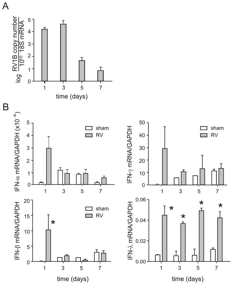
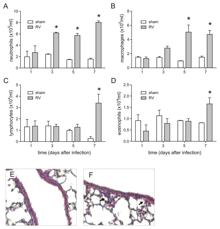
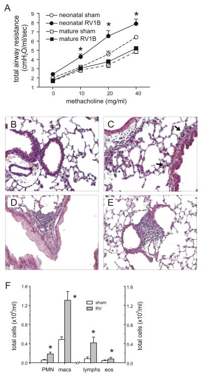
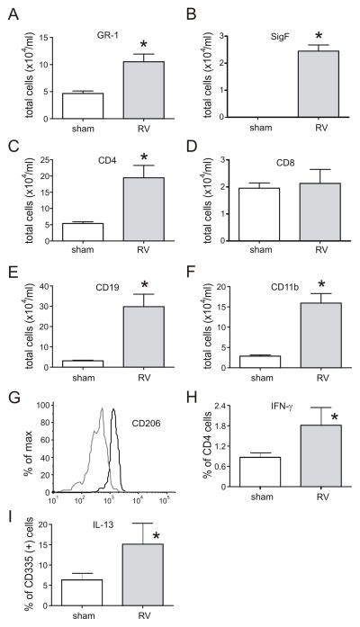
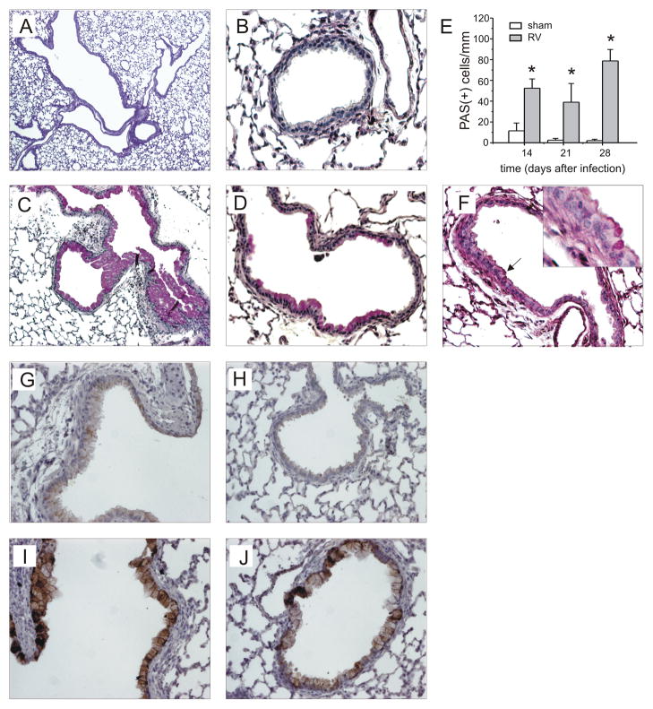
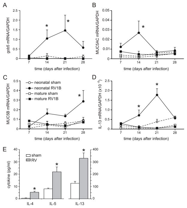
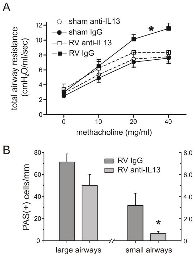
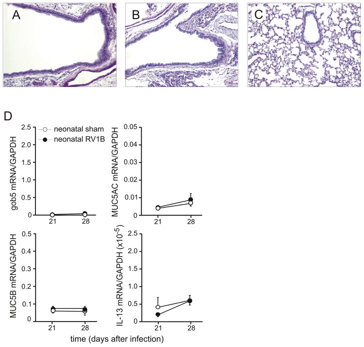
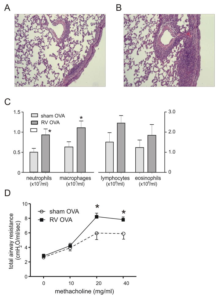
Recent studies link early rhinovirus (RV) infections to later asthma development. We hypothesized that neonatal RV infection leads to an IL-13-driven asthma-like phenotype in mice. BALB/c mice were inoculated with RV1B or sham on day 7 of life. Viral RNA persisted in the neonatal lung up to 7 d postinfection. Within this time frame, IFN-α, -β, and -γ peaked 1 d postinfection, whereas IFN-λ levels persisted. Next, we examined mice on day 35 of life, 28 d after initial infection. Compared with sham-treated controls, virus-inoculated mice demonstrated airways hyperresponsiveness. Lungs from RV-infected mice showed increases in several immune cell populations, as well as the percentages of CD4-positive T cells expressing IFN-γ and of NKp46/CD335(+), TCR-β(+) cells expressing IL-13. Periodic acid-Schiff and immunohistochemical staining revealed mucous cell metaplasia and muc5AC expression in RV1B- but not sham-inoculated lungs. Mucous metaplasia was accompanied by induction of gob-5, MUC5AC, MUC5B, and IL-13 mRNA. By comparison, adult mice infected with RV1B showed no change in IL-13 expression, mucus production, or airways responsiveness 28 d postinfection. Intraperitoneal administration of anti-IL-13 neutralizing Ab attenuated RV-induced mucous metaplasia and methacholine responses, and IL-4R null mice failed to show RV-induced mucous metaplasia. Finally, neonatal RV increased the inflammatory response to subsequent allergic sensitization and challenge. We conclude that neonatal RV1B infection leads to persistent airways inflammation, mucous metaplasia, and hyperresponsiveness, which are mediated, at least in part, by IL-13.


Figures

Figure 1
Figure 1. RV1B infection of neonatal mice elicits a brisk IFN response
Seven day-old BALB/c pups were intranasally infected with RV1B or sham control, and lungs harvested for analysis 1–7 days after infection. Positive-strand viral RNA (vRNA, panel A) and mRNAs encoding IFNs-α, -β and γ (B) were measured by qPCR. IFN expression was normalized by expression of the housekeeping gene GAPDH. N = 3–5 animals per time point, *different from sham, p<0.05, unpaired t test.
Figure 2
Figure 2. RV1B infection of neonatal mice induces airway inflammation
Seven day-old BALB/c pups were intranasally infected with RV1B or sham control, and lungs homogenates analyzed for inflammatory cell counts (A–D). Cytospins were stained with Diff-Quick. N = 3–4 animals per time point, *different from sham, p<0.05, unpaired t test. E, F. Images of lungs from sham- (E) and RV1B-infected (F) mice. Lungs were stained with hematoxylin and eosin. Arrows indicate focal areas of inflammation. These images are representative of three individual experiments of three mice per group.
Figure 3
Figure 3
RV1B infection of 7-day old BALB/c pups increases lung cytokine mRNA levels of CXCL1 (A), CXCL2 (B), TNF-α (C) and IL-13 (D). mRNA expression was measured by qPCR. N = 3–10 animals per time point, *different from sham, p<0.05, unpaired t test.
Figure 4
Figure 4
Neonatal RV infection induces persistent changes in airways responsiveness and inflammation. A. Effects of sham and RV1B infection of neonatal and mature mice on airway cholinergic responsiveness, measured 4 weeks after initial inoculation (day 35 of life for mice infected with RV as neonates and day 84 of life for mice infected with RV at maturity). Airway cholinergic responsiveness was assessed by measuring changes in total respiratory system resistance in response to increasing doses of nebulized methacholine. N = 6–10 animals per group, *different from sham-exposed mice of the same age, p<0.05, two-way ANOVA. B–E. Representative images of lungs harvested 4 weeks after neonatal sham- (B) or RV1B infection (C–E). Lungs were stained with hematoxylin and eosin. These images are representative of three individual experiments of three mice per group. F. Lung homogenate inflammatory cells from mice harvested 28 days after neonatal inoculation with sham- or RV. Cytospins were stained with Diff-Quick. N= 6 animals per group, *different from sham-exposed neonatal mice, p<0.05, unpaired t test.
Figure 5
Figure 5
Neonatal RV infection induces prolonged changes in airway inflammation. Cells from the minced lungs of 35 day-old mice infected with sham or RV at 7 days of age were analyzed by flow cytometry. Compared to sham-exposed mice, RV-infected mice showed significant increases in (A) Gr1-positive neutrophils and (B) Gr1-, SigF-double-positive and eosinophils, (C) CD4-positive T cells, (E) CD19-positive B cells and (F) CD11b-positive cells. There was no change in the number of CD8-positive cells (D). RV infection increased the CD206 expression of CD11b-positive cells (G; sham, grey line; RV, black line). Lung digests from RV-infected mice showed increases in the percentage of CD4-positive T cells expressing IFN-γ (H) and CD335-positive NK cells expressing IL-13 (I). N = 5–6 animals per group, *different from sham-exposed neonatal mice, p<0.05, rank sum test.
Figure 6
Figure 6
Neonatal RV infection causes airway mucous metaplasia. Lungs from mice inoculated with sham or RV1B at 7 days age were harvested on day 35 and stained with Periodic acid-Schiff (PAS). PAS staining in the large airways of sham-infected mice ranged from no signal to patches of dense staining (A). Sham-infected animals showed no staining in the small airways (B). In contrast, mice infected at day 7 of life showed some large airways which were completely filled with PAS-positive epithelial cells (C), as well as PAS-staining in the medium-sized and small airways (D). These images are representative of four individual experiments of 4–5 mice per group. E. PAS staining was quantified by using NIH ImageJ analysis software. RV-infected mice showed significantly increased PAS staining 14, 21 and 28 days after infection. N = 3 animals per group, *different from sham-exposed mice, p<0.05, unpaired t test. F. We also examined PAS staining 60 and 100 days after neonatal sham or RV treatment, on days 67 and 107 of life. RV-infected 67 day-old mice continued to show mucus (arrow, inset). These images are representative of two individual experiments of 4–5 mice per group. G–J. Lung tissues from sham-inoculated (G, H) and RV-infected mice (I, J) were also immunostained for MUC5AC. Signals were amplified and visualized using biotinylated anti-IgG and diaminobenzidine as a substrate. These images are representative of four individual experiments of 4–5 mice per group.
Figure 7
Figure 7
RV1B infection of 7-day old BALB/c pups induces persistent increases in the expression of mucus-related genes (A–C) and IL-13 (D). mRNA expression was measured by qPCR and normalized for GAPDH. N = 4 animals per time point, *different from sham-exposed mice of the same age, p<0.05, unpaired t test. E. IL-4, IL-5 and IL-13 protein levels from the lungs of RV-infected mice harvested three weeks after neonatal infection were significantly elevated compared to sham-exposed mice. Protein levels were measured by immune bioplex assay. (N = 4–5 animals per group, *different from sham-exposed mice, p<0.05, unpaired t test).
Figure 8
Figure 8
IL-13 neutralization partially attenuates RV-induced airways hyperresponsiveness and mucous cell metaplasia. A. Sham and RV1B-infected animals were treated with IgG or anti-IL-13. Airway cholinergic responsiveness was measured 4 weeks after initial inoculation. N = 4–6 animals per group, *different from sham-exposed mice, p<0.05, two-way ANOVA. B. PAS staining in the large and small airways of RV-infected mice treated with anti-IL13 or IgG. N = 6–8 animals per group, *different from sham-exposed mice, p<0.05, unpaired t test.
Figure 9
Figure 9
Effect of neonatal RV infection on IL-4R null mice. BALB/c-Il4ratm1Sz/J mice were inoculated with sham or RV at day 7 of life and airway tissues examined 28 days post-infection. Unlike RV-infected wild-type BALB/c mice (see Figure 6), IL-4R null mice showed no PAS-positive cells in large, medium or small airways (A–C). These images are representative of two individual experiments of three mice per group. D. Lung Gob5, MUC5AC, MUC5B and IL-13 mRNA expression was measured by qPCR. Unlike wild-type mice (see Figure 7), RV-infected IL-4R null mice failed to show an increase in mucus or IL-13 expression. N = 3–5 animals per group.
Figure 10
Figure 10
Effect of neonatal RV infection on the response to allergen sensitization and challenge. Mice were inoculated with sham or RV on day 7 of life, sensitized with intraperitoneal OVA on days 17 and 24 and challenged with intranasal OVA on days 32, 33 and 34. Compared to sham- and OVA-treated mice (panel A), RV-infected, OVA-treated mice showed greater peribronchial inflammation (B). Lung sections were stained with hematoxylin and eosin. Images represent two individual experiments of two mice per group. C, D. RV-infected mice also show greater OVA-induced bronchoalveolar neutrophils and macrophages (C) and airways cholinergic responsiveness (D). N = 3–6 animals per group, *different from sham, p<0.05, unpaired t test.

References

    1. Sigurs N, Bjarnason R, Sigurbergsson F, Kjellman B. Respiratory syncytial virus bronchiolitis in infancy is an important risk factor for asthma and allergy at age 7. Am J Respir Crit Care Med. 2000;161:1501–1507. - PubMed
    1. Sigurs N, Gustafsson PM, Bjarnason R, Lundberg F, Schmidt S, Sigurbergsson F, Kjellman B. Severe respiratory syncytial virus bronchiolitis in infancy and asthma and allergy at age 13. Am J Respir Crit Care Med. 2005;171:137–141. - PubMed
    1. Miller EK, Lu X, Erdman DD, Poehling KA, Zhu Y, Griffin MR, Hartert TV, Anderson LJ, Weinberg GA, Hall CB, Iwane MK, Edwards KM. Rhinovirus-associated hospitalizations in young children. J Infect Dis. 2007;195:773–781. - PMC - PubMed
    1. Lemanske RF, Jackson DJ, Gangnon RE, Evans MD, Li Z, Shult PA, Kirk CJ, Reisdorf E, Roberg KA, Anderson EL, Carlson-Dakes KT, Adler KJ, Gilbertson-White S, Pappas TE, Dasilva DF, Tisler CJ, Gern JE. Rhinovirus illnesses during infancy predict subsequent childhood wheezing. J Allergy Clin Immunol. 2005;116:571–577. - PubMed
    1. Jackson DJ, Gangnon RE, Evans MD, Roberg KA, Anderson EL, Pappas TE, Printz MC, Lee WM, Shult PA, Reisdorf E, Carlson-Dakes KT, Salazar LP, DaSilva DF, Tisler CJ, Gern JE, Lemanske RF., Jr Wheezing rhinovirus illnesses in early life predict asthma development in high-risk children. Am J Respir Crit Care Med. 2008;178:667–672. - PMC - PubMed

Publication types

MeSH terms