. 2012 Jul-Aug;26(6):556-69.
doi: 10.1177/1545968311421934. Epub 2012 Feb 13.

Methods for functional assessment after C7 spinal cord hemisection in the rhesus monkey

Affiliations

Methods for functional assessment after C7 spinal cord hemisection in the rhesus monkey

Yvette S Nout et al. Neurorehabil Neural Repair. 2012 Jul-Aug.

Abstract

Background: Reliable outcome measures are essential for preclinical modeling of spinal cord injury (SCI) in primates.

Measures: need to be sensitive to both increases and decreases in function in order to demonstrate potential positive or negative effects of therapeutics.

Objectives: To develop behavioral tests and analyses to assess recovery of function after SCI in the nonhuman primate.

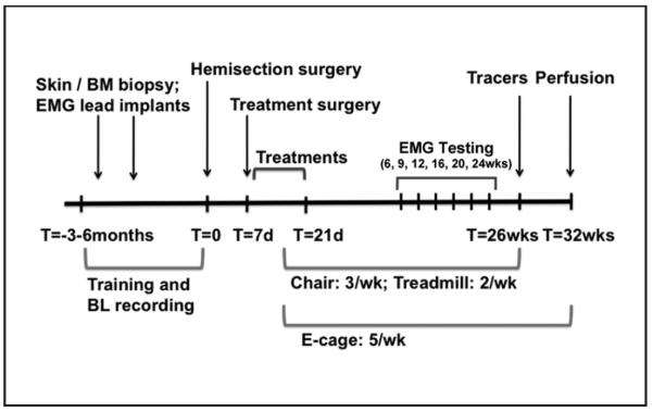
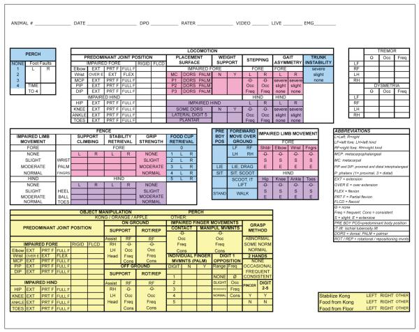
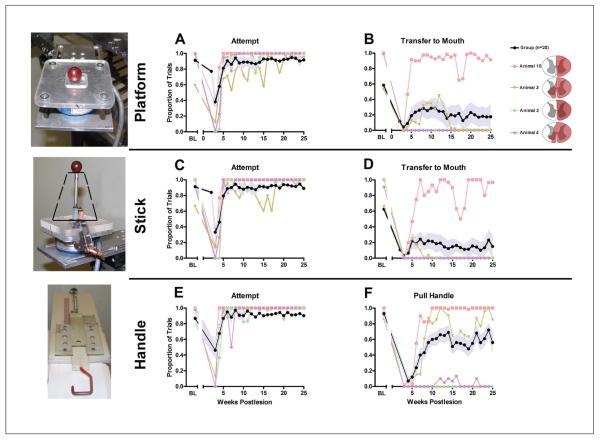
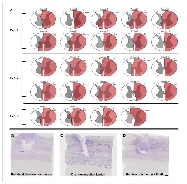
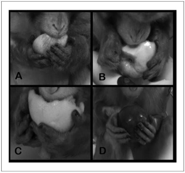
Methods: In all, 24 male rhesus macaques were subjected to complete C7 lateral hemisection. The authors scored recovery of function in an open field and during hand tasks in a restraining chair. In addition, EMG analyses were performed in the open field, during hand tasks, and while animals walked on a treadmill. Both control and treated monkeys that received candidate therapeutics were included in this report to determine whether the behavioral assays were capable of detecting changes in function over a wide range of outcomes.

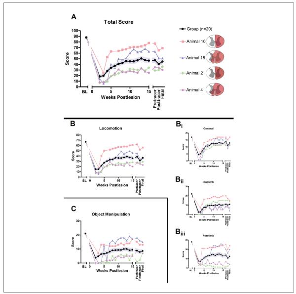
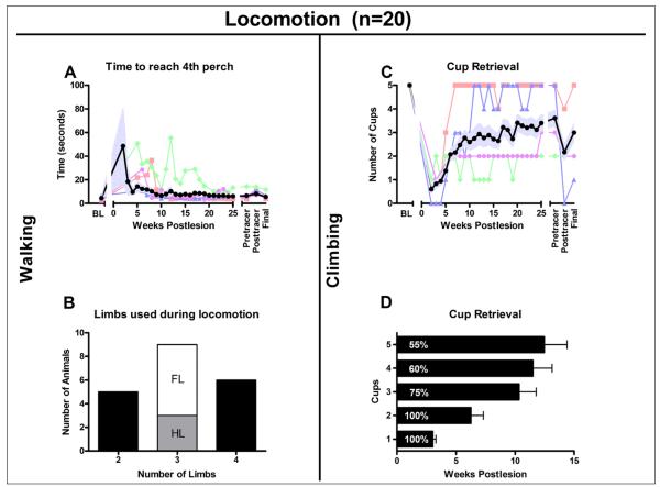
Results: The behavioral assays are shown to be sensitive to detecting a wide range of motor functional outcomes after cervical hemisection in the nonhuman primate. Population curves on recovery of function were similar across the different tasks; in general, the population recovers to about 50% of baseline performance on measures of forelimb function.

Conclusions: The behavioral outcome measures that the authors developed in this preclinical nonhuman primate model of SCI can detect a broad range of motor recovery. A set of behavioral assays is an essential component of a model that will be used to test efficacies of translational candidate therapies for SCI.


Figures

Figure 1
Figure 1
Timeline: T = 0 indicates the time of spinal cord hemisection. In a period of 3 to 6 months before spinal cord hemisection, animals underwent 2 preparatory surgeries: a skin or bone marrow (BM) biopsy and implantation of EMG leads. In addition, animals were trained to perform chair, treadmill, and open-field tasks. Training and collection of baseline (BL) data on these tasks occurred up until the week of spinal cord hemisection. Then, 7 days after spinal cord hemisection, animals underwent a treatment surgery followed by 14 days of treatment delivery in the hospital and home cage. Behavioral testing in the chair (3 times weekly), treadmill (twice weekly), and open field (5 times weekly) resumed once animals were ready (during week 2 after spinal cord hemisection) and continued until week 25 (chair and treadmill) and week 32 (open field). EMG and 3D video recording sessions occurred before the lesion and at weeks 6, 9, 12, 16, 20, and 24. Animals underwent surgery 6 weeks prior to perfusion for delivery of tracers into the motor cortex, brainstem, and spinal cord.
Figure 2
Figure 2
Open field data collection form: This is the data form used to record function in the open field. Data were used to generate an overall total recovery score (Figures 3 and 4). This total score comprised a locomotion and object manipulation subscore (yellow). The locomotion score was generated from a general subscore (blue), a forelimb subscore (pink), and a hind limb subscore (purple).
Figure 3
Figure 3
Open-field analysis (n = 20): A. The overall total score for behavior assessed in the open field over time (maximum score = 88) for a group of 20 animals. The group mean ± standard error of the mean (SEM) is shown in black and light blue, respectively, and 4 representative individual subject recovery curves are also shown; schematic lesion reconstructions for these subjects are illustrated on the right. Immediately after hemisection, function is substantially reduced but shows recovery over time, although significant deficits remain. The total score comprises the locomotion score (B; maximum = 67) and the object manipulation score (C; maximum = 21). Similar to the total score, after hemisection, specific aspects of locomotor function are lost but show varying degrees of recovery (B). The subdivisions of the locomotion score (B), including a general subscore (Bi; maximum = 17), a hind limb subscore (Bii; maximum = 22), and a forelimb subscore (Biii; maximum = 28), all show loss of function followed by recovery similar to the overall locomotion and total scores. Group recovery on the general scale (Bi) was approximately 70% of baseline. This measured overall ability to independently move through the cage and retrieve food items. Specific methods, for example, ipsilateral hand participation in these maneuvers, are not taken into account in this subscore, and performance on this subcomponent showed that most animals recovered their ability to perform the tasks in the open field fairly well. Hind limb (Bii) and forelimb (Biii) functions recover to approximately 50% and 45% of baseline, respectively. On all graphs, the group mean ± SEM (n = 20) is shown in black and pale blue, respectively, and 4 individual recovery curves selected to illustrate the full range are also shown (as are their lesion reconstructions on the right). The 4 selected individuals are the same for Figures 3 and 4 and were selected to illustrate the range of recoveries. Abbreviation: BL, baseline.
Figure 4
Figure 4
Recovery of walking (A, B) and climbing (C, D), n = 20. A. Time required to reach the fourth perch after entering the open field. Immediately after surgery, the time to fourth perch was prolonged, but animals recovered rapidly to achieve times that were just slightly longer than at baseline. Animals used 2, 3, or all 4 limbs for locomotion. B. The number of animals that use 2, 3 (white: using right forelimb; gray: using right hind limb), or all 4 limbs for walking. C and D, food retrieval from the cups. C. Mean number of cups emptied. D. Percentage of animals able to reach cups 1 to 5 and the average time ± standard error of the mean (SEM) they took to accomplish this. In A and C the group mean ± SEM (n = 20) is shown in black and pale blue, and 4 individual recovery curves are also shown. The 4 selected individuals are the same for Figures 3 and 4 and were selected to illustrate the range of recoveries. In D, the group mean ± SEM is shown. Abbreviation: BL, baseline.
Figure 5
Figure 5
Recovery of function over time on skilled forelimb tasks (n = 20): performance on tasks of skilled forelimb use while seated in a restraining chair is shown on 3 tasks: object retrieval from a flat platform (upper panel), grape retrieval from a vertical stick (middle panel, funnel position indicated with black dashed line), and a handle-pull task (lower panel), with an example of each task apparatus shown at the left. A, C, E: The proportion of trials on which the animals attempted food retrieval (Attempt) on each chair-based task, whether successful or not. In the 4-week period after hemisection, few animals attempted to perform the task. Rapid recovery in ability to initiate movement is observed by week 5. B, D, F: Successful completions of platform task (food retrieval and placement in mouth). B. Stick task (food retrieval from vertical stick and placement in mouth). D. Handle task (pull handle). F. Group means ± standard error of the mean are shown in black and pale blue, respectively, and 4 individual recovery curves in color (individual lesion reconstructions are shown on the right). Three of the 4 selected individuals are the same for Figures 3, 4, and 5. A range of initial deficits are evident, with some followed by substantial recovery (eg, animals 3 and 10). Abbreviation: BL, baseline.
Figure 6
Figure 6
Spasticity assessment on the Ashworth Scale (n = 16): assessment of rigidity of the right forelimb using the Ashworth Scale. An increase in hand/arm spasticity was observed in most animals during the course of recovery. Wide variability was present, with some animals developing little increase in spasticity and occasional animals developing spastic distal forelimbs. Group means ± standard error of the mean are shown in black and pale blue, respectively.
Figure 7
Figure 7
Lesions produced by hemisection (n = 24): A. Reconstructions of the lesion site for all animals described in this communication; red indicates lesioned area. In 4 animals, small areas of the right hemicord were spared. In 5 animals, there was a complete lesion of the right hemicord with no obvious damage to the left hemicord. The remaining 15 animals had complete lesions with varying degrees of left hemicord damage. B, C, and D. Nissl-stained horizontal sections through the lesion for 3 selected subjects. The lesions depicted include a complete hemisection that extended to the midline (B), an overhemisection that extended over the midline into the contralateral side (C), and a hemisection lesion with a treatment graft in the lesion site (D). In animals receiving a cellular transplant (D), the lesion site was generally completely filled with cells, likely a combination of transplanted and endogenous cells. Dashed line indicates midline in B, C, and D. Scale bar = 500 μm.
Figure 8
Figure 8
Range of recovery of hand function for object manipulation: Animals eating an orange or apple are shown. A. An animal prior to hemisection (baseline); both hands are used to manipulate the apple. Palmar hand and digit contact is present as is a normal distance between digits. Wrist and digital movements (not visible in this still image) are present. B. An example of poorly recovered hand function. The right hand is clubbed and the object is supported against the dorsal aspect of the digits. C. An animal using a slightly more normal grasping method to manipulate the orange; however, full palmar contact does not occur. D. An animal that regained a near-normal grasping method; note, however, that there is still no distance between digits 1 and 2. This lack of interdigital space and movement of digit 1 appear to be one of the last components of grasping to recover after the lesion. Also note the position of digit 1 on the normal left hand and digit 1 on the ipsilesional right hand in each picture.

Similar articles

Cited by

References

    1. Turner WA. On hemisection of the spinal cord. Brain. 1891;14:496–522.
    1. Denny-Brown D. The Cerebral Control of Movement. Liverpool University Press; Liverpool, UK: 1966.
    1. Mettler FA. Observations on the consequences of large, subtotal lesions of the simian spinal cord. J Comp Neurol. 1944;81:339–360.
    1. Mettler FA, Liss H. Functional recovery in primates after large subtotal spinal cord lesions. J Neuropathol Exp Neurol. 1959;18:509–516.
    1. Brinkman J, Bush BM, Porter R. Deficient influence of peripheral stimuli on precentral neurones in monkeys with dorsal column lesions. J Physiol. 1978;276:27–48. - PMC - PubMed

Publication types

MeSH terms