. 2012 Jan 15;125(Pt 2):399-410.
doi: 10.1242/jcs.089797. Epub 2012 Feb 13.

CoREST acts as a positive regulator of Notch signaling in the follicle cells of Drosophila melanogaster

Affiliations

CoREST acts as a positive regulator of Notch signaling in the follicle cells of Drosophila melanogaster

Elena Domanitskaya et al. J Cell Sci. .

Abstract

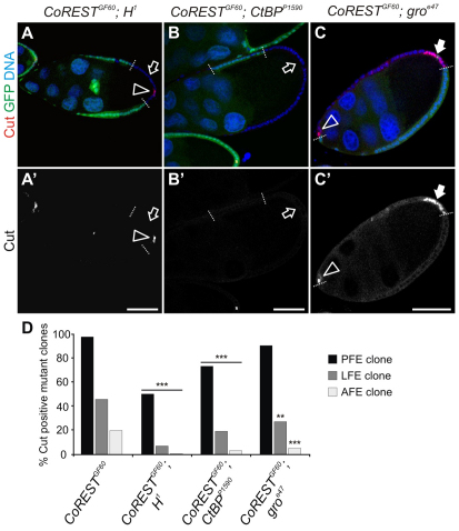
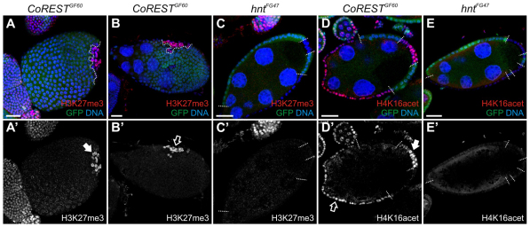
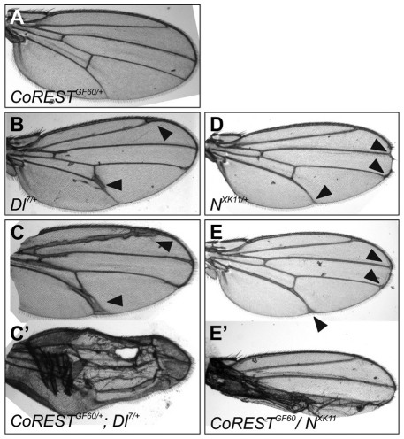
The Notch signaling pathway plays important roles in a variety of developmental events. The context-dependent activities of positive and negative modulators dramatically increase the diversity of cellular responses to Notch signaling. In a screen for mutations affecting the Drosophila melanogaster follicular epithelium, we isolated a mutation in CoREST that disrupts the Notch-dependent mitotic-to-endocycle switch of follicle cells at stage 6 of oogenesis. We show that Drosophila CoREST positively regulates Notch signaling, acting downstream of the proteolytic cleavage of Notch but upstream of Hindsight activity; the Hindsight gene is a Notch target that coordinates responses in the follicle cells. We show that CoREST genetically interacts with components of the Notch repressor complex, Hairless, C-terminal Binding Protein and Groucho. In addition, we demonstrate that levels of H3K27me3 and H4K16 acetylation are dramatically increased in CoREST mutant follicle cells. Our data indicate that CoREST acts as a positive modulator of the Notch pathway in the follicular epithelium as well as in wing tissue, and suggests a previously unidentified role for CoREST in the regulation of Notch signaling. Given its high degree of conservation among species, CoREST probably also functions as a regulator of Notch-dependent cellular events in other organisms.


Figures

Fig. 1.
Fig. 1.
GF60 function is required for the M/E switch. Egg chambers at stage 10 (A) and 8 (B,C) of oogenesis containing GF60 mutant follicle cell clones, stained with Hoechst (A,A′,B,C) to label DNA, F-actin (A), PH3 (B,B′) and CycB (C,C′). Mutant cells are identifiable by the absence of GFP (green), the borders between mutant clones and neighboring wild-type cells are marked by dotted lines. Scale bars: 25 μm. (A,A′) GF60 mutant follicle cell nuclei (filled arrow in A′) are smaller and more crowded than neighboring wild-type cell nuclei (open arrows in A′). (B,B′) PH3-positive cells (filled arrow in B′) are observed in the GF60 mutant PFCs after stage 7, indicating continued mitotic division. (C,C′) CycB is continuously expressed in the GF60 mutant PFCs (filled arrow in C′) but is not observed in neighboring wild-type cells (open arrow in C′) in an egg chamber at stage 9.
Fig. 2.
Fig. 2.
The defect in the M/E switch in GF60 is caused by a mutation in CoREST. (A) Schematic representation of the intron–exon structure of CoREST. White rectangles represent the 5′ and 3′ UTR, grey rectangles represent translated regions. The third intron is mutated in the GF60 allele leading to the substitution of A for G (highlighted in red), which abolishes its splicing (data not shown). The stop codon in the third intron is highlighted in yellow. (B) Schematic diagrams illustrating three Drosophila CoREST splice variants [adopted from Dallman et al. (Dallman et al., 2004)]. ELM2 domains are depicted as green rectangles, SANT domains are represented by blue ovals. The short, medium and long isoforms of CoREST are 273, 590 and 824 aa, respectively. The GF60 mutation leads to the truncation of CoREST before the ELM2 domains, and is indicated by a red star. (C) Stage 7 egg chamber containing CoRESTGF60 homozygous mutant clones stained with anti-CoREST antibodies (red in C, white in C′) and Hoechst (blue in C) to label DNA. Mutant clones are identifiable by the absence of GFP (green in C, white in C″), and the borders between mutant clones and neighboring wild-type cells are marked by dotted lines (C′,C″). CoREST is present in wild-type follicle epithelial cells (open arrow in C′), and absent from mutant clones (filled arrow in C′). Egg chambers are oriented with the posterior to the right. Scale bar: 50 μm.
Fig. 3.
Fig. 3.
CoREST function is required for Notch signaling in the follicle epithelial cells. Egg chambers stained with Hoechst (A–F) to label nuclei, and with anti-Hnt (A–C) and anti-Cut (D–F) antibodies. CoRESTGF60 homozygous mutant clones are identifiable by the absence of GFP. The borders between mutant clones and neighboring wild-type cells are marked by dotted lines (E,F,E′,F′). Egg chambers are oriented with the posterior to the right. Scale bars: 50 μm. (A) In the wild-type FCs Hnt is upregulated after stage 7 of oogenesis (open arrows, A′). (B) Egg chamber containing whole follicle-cell clone. Hnt fails to be upregulated after stage 7 in CoRESTGF60 homozygous mutant PFCs (filled arrow, B′), whereas Hnt is normally upregulated after stage 7 in CoRESTGF60 homozygous mutant lateral and anterior FCs (open arrow, B′). (C) Absence of Hnt expression is observed in CoRESTGF60 homozygous mutant PFCs and lateral FCs in the Dl7 heterozygous background (filled arrows, C′), due to the increased penetrance of the mutant phenotype. Hnt expression is indicated by the open arrow in C′. (D) Notch activation leads to the downregulation of Cut expression after stage 7 in entire follicle epithelium except for anterior and posterior polar cells (open arrowheads, D′). (E) Cut is continuously present after stage 7 in the CoRESTGF60 homozygous mutant PFCs (filled arrow, E′), but it is correctly downregulated after stage 7 in CoRESTGF60 homozygous mutant lateral and anterior FCs (open arrow, E′). (F) Cut expression is present in the CoRESTGF60 homozygous mutant posterior, lateral and anterior follicle cells in the Dl7 heterozygous background (filled arrows, F′), which indicates an increased penetrance of the mutant phenotype.
Fig. 4.
Fig. 4.
CoREST acts downstream of Notch proteolytic cleavage and upstream of Hnt in the regulation of Notch signaling and the M/E switch. CoRESTGF60 homozygous mutant clones positively marked by GFP; the borders between mutant clones and neighboring wild-type cells are marked by dotted lines. Egg chambers are oriented with the posterior to the right. Scale bars: 20 μm. (A–C′) Egg chambers, at stage 8 to stage 9, containing CoRESTGF60 homozygous mutant clones with simultaneous overexpression of NFL (A,A′), NEXT (B,B′) or NICD (C,C′), and stained with an antibody against Cut (red in A–C, white in A′–C′). In CoRESTGF60 homozygous mutant clones with simultaneous expression of either of the Notch cleavage products, Cut was continuously expressed in PFCs (filled arrows in A′,B′,C′). (D,E″) Egg chambers, at stage 4 to stage 5, containing CoRESTGF60 homozygous mutant clones with simultaneous Hnt misexpression, stained either with anti-Hnt antibodies (red in D, white in D′) or anti-Cut antibodies (red in E, white in E′), and with Hoechst to indicate the nuclei (blue in D and E and white in D″and E″). The hntEP55 allele contains the UAS sequence inserted 585 bp upstream of the hnt gene and is used for Hnt misexpression. Misexpression of Hnt in CoRESTGF60 homozygous mutant clones (open arrow in D′) is sufficient to downregulate Cut before stage 6 (open arrow in E′) and to drive follicle cells to enter endocycle prematurely (open arrow in E″).
Fig. 5.
Fig. 5.
Notch mutant phenotype in CoRESTGF60 mutants is suppressed in H1, CtBPP150 and groe47 heterozygous backgrounds. Egg chambers containing GF60 mutant follicle cell clones stained with Hoechst (A–C) to label DNA, and anti-Cut antibody (A–C, A′–C′). Mutant cells are identifiable by the absence of GFP (green), the borders between mutant clones and neighboring wild-type cells are marked by dotted lines. Egg chambers are oriented with the posterior to the right. Scale bars: 50 μm. (A,B) In egg chambers containing CoRESTGF60 homozygous mutant follicle cell clones in the H1 heterozygous background (A) or in the CtBPP150 heterozygous background (B), the Notch phenotype was strongly suppressed as indicated by complete Cut downregulation in the mutant clones after stage 7 (open arrow in A,A′,B,B′), except for the polar cells (open arrowhead in A,A′). (C) In egg chambers containing CoRESTGF60 homozygous mutant clones in the groe47 heterozygous background, Cut was continuously expressed in PFCs after stage 7 (filled arrow in C′), and the strength of this phenotype was similar to the phenotype in CoRESTGF60 homozygous mutant FC clones in the wild-type background. Cut staining in the polar cells is indicated by open arrowhead (C,C′). (D) Percentage of the egg chambers with Cut-positive cells in the CoRESTGF60 homozygous mutant posterior (black), lateral (dark gray) and anterior (light gray) FC clones, in the control, H1, CtBPP150 or Groe47 heterozygous backgrounds. Percentage of egg chambers with Cut-positive cells in CoRESTGF60 homozygous mutant clones in wild-type background: 97.5% of PFC clones (n=193), 46% of lateral clones (n=218), 20% of anterior clones (n=110); in CoRESTGF60 homozygous mutant clones in the H1 heterozygous background: 51% of PFC clones (n=73), 6% of lateral clones (n=64), 0% of anterior clones (n=52); in CoRESTGF60 homozygous mutant clones in the CtBPP150 heterozygous background: 73% of PFC clones (n=224), 19% of lateral clones (n=206), 3% of anterior clones (n=135); in CoRESTGF60 homozygous mutant clones in the groe47 heterozygous background: 90% PFC clones (n=73), 27% lateral clones (n=55), 5% anterior clones (n=38). The χ2 test was applied for statistical analysis. ***P<0.001 and **P<0.01 for the comparison between CoRESTGF60 homozygous mutant clones in the wild-type background and all other groups.
Fig. 6.
Fig. 6.
Loss of CoREST alters global levels of chromatin methylation and acetylation. Egg chambers stained with Hoechst to label nuclei (blue in A–E), and antibodies specific for H3K27me3 (red in A–C, white in A″–C″), and H4K16 acetylation (red in D and E, white in D″ and E″). CoRESTGF60 homozygous mutant clones (A,B,D) and hntFG47 homozygous mutant clones (C,E) are identifiable by the absence of GFP. The borders between mutant clones and neighboring wild-type cells are marked by dotted lines. Egg chambers are oriented with the posterior to the right. Scale bars: 20 μm. (A,B) A higher level of H3K27me3 staining was observed in CoRESTGF60 homozygous mutant FC clones, independent of clone position (filled arrow indicates posterior clone in A′, open arrow indicates lateral clone in B′) in stage 7 and older egg chambers. (C) The level of H3K27me3 staining in hntFG47 homozygous mutant follicle epithelial was comparable to that in wild-type neighboring cells. (D) The level of H4K16 acetylation was higher in CoRESTGF60 homozygous mutant follicle cell clones independent of clone position (filled arrow indicates a posterior clone in D′, and open arrow indicates an anterior-lateral clone in D′) in the stage 7 and older egg chambers. (C) The level of H4K16 acetylation was comparable in hntFG47 homozygous mutant follicle epithelial cells with their wild-type neighboring cells.
Fig. 7.
Fig. 7.
CoREST genetically interacts with Notch and Delta in wing development. The CoRESTGF60 mutant can modify the phenotype of Delta and Notch alleles in transheterozygotes. (A) A wing from a female heterozygous for CoRESTGF60 shows wild-type wing morphology. (B) Dl7/+. (C) CoRESTGF60/+; Dl7/+. The CoRESTGF60 mutation enhances the phenotype of the wing vein thickening (filled arrowheads) and the delta between L4 and L5. (D) NXK11/+. (E) CoRESTGF60/NXK11. The CoRESTGF60 mutation also enhances the thickening of deltas (filled arrowheads). In addition, wing blistering is present in some instances (C′,E′).

Similar articles

Cited by

References

    1. Abrajano J. J., Qureshi I. A., Gokhan S., Zheng D., Bergman A., Mehler M. F. (2009a). Differential deployment of REST and CoREST promotes glial subtype specification and oligodendrocyte lineage maturation. PLoS ONE 4, e7665 - PMC - PubMed
    1. Abrajano J. J., Qureshi I. A., Gokhan S., Zheng D., Bergman A., Mehler M. F. (2009b). REST and CoREST modulate neuronal subtype specification, maturation and maintenance. PLoS ONE 4, e7936 - PMC - PubMed
    1. Abrajano J. J., Qureshi I. A., Gokhan S., Molero A. E., Zheng D., Bergman A., Mehler M. F. (2010). Corepressor for element-1-silencing transcription factor preferentially mediates gene networks underlying neural stem cell fate decisions. Proc. Natl. Acad. Sci. USA 107, 16685-16690 - PMC - PubMed
    1. Andres M. E., Burger C., Peral-Rubio M. J., Battaglioli E., Anderson M. E., Grimes J., Dallman J., Ballas N., Mandel G. (1999). CoREST: a functional corepressor required for regulation of neural-specific gene expression. Proc. Natl. Acad. Sci. USA 96, 9873-9878 - PMC - PubMed
    1. Artavanis-Tsakonas S., Muskavitch M. A. (2010). Notch: the past, the present, and the future. Curr. Top. Dev. Biol. 92, 1-29 - PubMed

Publication types

MeSH terms

LinkOut - more resources