. 2012 Feb 1;125(Pt 3):673-84.
doi: 10.1242/jcs.091355. Epub 2012 Feb 13.

Epithelial organization and cyst lumen expansion require efficient Sec13-Sec31-driven secretion

Affiliations

Epithelial organization and cyst lumen expansion require efficient Sec13-Sec31-driven secretion

Anna K Townley et al. J Cell Sci. .

Abstract

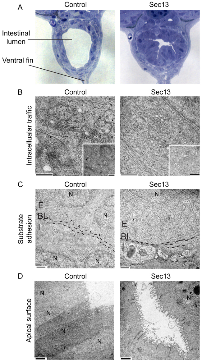
Epithelial morphogenesis is directed by interactions with the underlying extracellular matrix. Secretion of collagen and other matrix components requires efficient coat complex II (COPII) vesicle formation at the endoplasmic reticulum. Here, we show that suppression of the outer layer COPII component, Sec13, in zebrafish embryos results in a disorganized gut epithelium. In human intestinal epithelial cells (Caco-2), Sec13 depletion causes defective epithelial polarity and organization on permeable supports. Defects are seen in the ability of cells to adhere to the substrate, form a monolayer and form intercellular junctions. When embedded in a three-dimensional matrix, Sec13-depleted Caco-2 cells form cysts but, unlike controls, are defective in lumen expansion. Incorporation of primary fibroblasts within the three-dimensional culture substantially restores normal morphogenesis. We conclude that efficient COPII-dependent secretion, notably assembly of Sec13-Sec31, is required to drive epithelial morphogenesis in both two- and three-dimensional cultures in vitro, as well as in vivo. Our results provide insight into the role of COPII in epithelial morphogenesis and have implications for the interpretation of epithelial polarity and organization assays in cell culture.


Figures

Fig. 1.
Fig. 1.
Disorganization and limited expansion of the zebrafish gut lumen following Sec13 suppression. (A) Transverse sections through the gut of 5 dpf zebrafish embryos injected with control (left) or Sec13 morpholinos (right). Sections (1 μm) were taken from a comparable level along the cranio–caudal axis of age-matched embryos. Note the limited expansion of the lumen of the gut and disorganization of the gut epithelium in Sec13 morphants. (B) The intracellular organization of the ER–Golgi interface is disrupted in morphant epithelial cells. Suppression of Sec13-induced distension of the ER and accumulation of budding profiles (see insets for second example). (C) Sec13-depleted intestinal epithelial cells are attached to surrounding cells but a clear basal lamina (indicated by dotted lines, compare with control) is missing. (D) TEM of intestinal epithelial cells reveals that the microvilli seam was intact in Sec13 morphants but cell monolayer formation was disorganized, as evident from the diverse range of surface areas of the cells reaching the lumen. Total of three embryos analyzed for each oligo used. The intestinal phenotype of all of zebrafish included in this study was defined by showing pectoral fin defects as published previously (Townley et al., 2008). BL, basal lamina; E, epithelial cells; I, intestinal lining; N, nucleus. Scale bars: 0.5 μm (B,C); 1 μm (D).
Fig. 2.
Fig. 2.
Stable suppression of Sec13 expression in Caco-2 cells. (A) Immunoblotting of lysates from cells transduced with lentiviral constructs as indicated. Lysates of cells stably expressing shRNA were immunoblotted for Sec13, Sec31A and α-tubulin as indicated. Molecular markers are shown in kDa. The asterisk marks a nonspecific band detected by the antibody. (B) X–Z reconstructions of DAPI-labelled nuclei of epithelial monolayers grown for 14 days on a permeable support from control or Sec13-1 Caco-2 cells. (C) Automatic detection of cell nuclei and pseudo-colouring highlights the disorganization of the epithelial layer following suppression of Sec13. (D) TEM of polarized Caco-2 monolayer cultures from control and Sec13-1-suppressed cells. Note the extensive interdigitation of intercellular junctions on suppression of Sec13 (arrows). Scale bars: 2 μm.
Fig. 3.
Fig. 3.
Electron microscopy of epithelial organization in Sec13-depleted Caco-2 cells. (A,B) SEM of microvilli on the apical surface of Caco-2 monolayers grown for 14 days on transwell filters: (A) control cells, (B) Sec13-depleted cells. (C–F) Polarized Caco-2 cells stably expressing control or Sec13 shRNAs as indicated were examined for (C) polarity (i.e. the formation of monolayers), (D) attachment to the substratum (dotted lines indicate basal cell surface), (E) intercellular adhesion (cell–cell attachment indicated by arrows) and (F) ER–Golgi membrane organization. (G–J) Cells were also examined by TEM following growth for 14 days on collagen-IV-coated permeable supports (G, polarity; H, attachment; I, adhesion; J, ER and Golgi membrane traffic). Two independent samples were analyzed for each condition. N, nuclei. Scale bars: 2 μm (A–C); 1 μm (D,E); 0.5 μm (F).
Fig. 4.
Fig. 4.
Sec13 suppression disrupts cyst formation by Caco-2 cells embedded in 3D matrix. (A) Formation of cysts by Caco-2 cells grown for 7 days in 3D matrix is inhibited by Sec13 suppression. Organization of the cell monolayer is affected compared with controls (DAPI labelling of nuclei), as is expansion of the central lumen (highlighted by phalloidin labelling of filamentous actin). Cysts are also smaller. (B) Basolateral targeting is unaffected as shown by labelling for ESA. (C) The number of cells per cyst is also decreased, indicating a cell division defect. (D) Quantification shows that lumen size (lumen area at the point of maximum cyst width) is significantly decreased on Sec13 suppression (with or without the presence of 6-Bnz). In A–D, n>40 cysts in each of three independent experiments performed for each experimental condition. Error bars represent s.e.m. P values compare depleted samples with control. (E) Alignment of the mitotic spindle (tubulin labelling) is perturbed in Sec13-depleted cysts, with frequent mitotic profiles seen in the centre of cysts as well as in the limiting layer. (F) Quantification of spindle alignment in these cysts. Median values are shown by the horizontal bar within each box; boxes show 25th and 75th percentiles; whiskers show the spread of the data. In E and F, n=30 cysts for each shRNA from three independent experiments. Scale bars: 20 μm.
Fig. 5.
Fig. 5.
Deposition and assembly of ECM surrounding epithelial cysts is impaired following Sec13 depletion. (A,B) Collagen I and perlecan deposition by cells grown on glass coverslips was analyzed by immunofluorescence microscopy following removal of cells. B shows quantification of results. (C,D) Collagen IV assembly around epithelial cysts grown in 3D matrix is unaffected by Sec13 suppression. By contrast, laminin-1 labelling around control cysts covers a much greater area than that surrounding cysts formed from Sec13-depleted cells. The intensity of labelling immediately surrounding the cyst is also significantly reduced following Sec13 suppression. Three examples are shown in C. Quantification of laminin labelling is shown in D. Median values are shown by the horizontal bar within each box; boxes show 25th and 75th percentiles; whiskers show the spread of the data. n=26 cysts total from three independent experiments. (E) Immunoblotting of cell-derived matrices. Cells were grown for 10 days in tissue culture dishes with ascorbic acid feeding. Cells were denuded and the remaining matrix was removed and added to sample buffer. Samples were immunoblotted with the following antibodies: laminin γ-2, laminin β-3 and rabbit polyclonal antibody against laminin (which recognises A chain, B1 chain and B2 chain). Molecular mass is indicated in KDa. Scale bars: 20 μm.
Fig. 6.
Fig. 6.
Cyst morphogenesis defects in 3D matrix following Sec13 suppression can be substantially rescued by co-culture with human fibroblasts. (A) Lumen formation is partially restored in cysts formed from Sec13-suppressed cells in the presence of fibroblasts. Cysts are more spherical, showing well-defined apical actin organization. Cells are still seen within the cyst in addition to the limiting monolayer, suggesting a partial but incomplete recovery of morphogenesis. Scale bar: 20 μm. (B) Quantification of cyst lumen area from three independent experiments shows that growth in the presence of fibroblasts restored lumen expansion to Sec13-1 depleted cysts. Similar results were obtained for Sec13-2 cells. Boxes show the median with 25th and 75th percentiles; whiskers show the spread of the data. n>20 (typically 20–30) cysts in each of three independent experiments performed for each experimental condition. Results of statistical testing (Student's t-test) compared with controls in 3D matrix alone are indicated.
Fig. 7.
Fig. 7.
Rescue of Sec13 lumen defect by addition of fibroblast-conditioned medium. Caco-2 cells expressing scrambled shRNA or Sec13 shRNA were embedded in Geltrex and grown for 7 days. Cells were grown in the presence of either control (Caco-2) medium or conditioned medium taken from a confluent dish of NDF. (A) Scramble shRNA-transfected Caco-2 cells plus Caco-2 medium. (B) Scramble shRNA-transfected Caco-2 cells plus NDF medium. (C) Sec13 shRNA-transfected Caco-2 cells plus Caco-2 medium. (D) Sec13 shRNA-transfected Caco-2 cells plus NDF medium. (E–H) Representation of mean lumen area for cysts formed in A–D; green represents basolateral surface with the apical surface in red. (I) Quantification of mean lumen size for cysts in A–D; median values are shown by the horizontal bar within each box; boxes show 25th and 75th percentiles; whiskers show the spread of the data. n>20 cysts in each of three independent experiments performed for each experimental condition. Scale bars: 10 μm.

Similar articles

Cited by

References

    1. Abrams E. W., Andrew D. J. (2005). CrebA regulates secretory activity in the Drosophila salivary gland and epidermis. Development 132, 2743-2758 - PubMed
    1. Antonny B., Madden D., Hamamoto S., Orci L., Schekman R. (2001). Dynamics of the COPII coat with GTP and stable analogues. Nat. Cell Biol. 3, 531-537 - PubMed
    1. Antonny B., Gounon P., Schekman R., Orci L. (2003). Self-assembly of minimal COPII cages. EMBO Rep. 4, 419-424 - PMC - PubMed
    1. Aridor M., Bannykh S. I., Rowe T., Balch W. E. (1999). Cargo can modulate COPII vesicle formation from the endoplasmic reticulum. J. Biol. Chem. 274, 4389-4399 - PubMed
    1. Barlowe C., Orci L., Yeung T., Hosobuchi M., Hamamoto S., Salama N., Rexach M. F., Ravazzola M., Amherdt M., Schekman R. (1994). COPII: a membrane coat formed by Sec proteins that drive vesicle budding from the endoplasmic reticulum. Cell 77, 895-907 - PubMed

Publication types

MeSH terms