. 2012 Feb 1;125(Pt 3):614-24.
doi: 10.1242/jcs.089367. Epub 2012 Feb 13.

Differential regulation of actin microfilaments by human MICAL proteins

Affiliations

Differential regulation of actin microfilaments by human MICAL proteins

Sai Srinivas Panapakkam Giridharan et al. J Cell Sci. .

Abstract

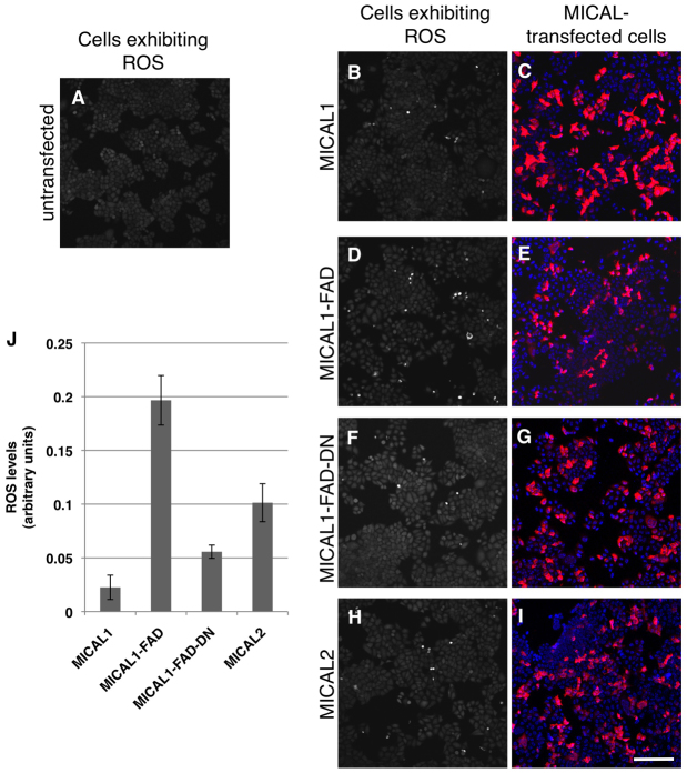
The Drosophila melanogaster MICAL protein is essential for the neuronal growth cone machinery that functions through plexin- and semaphorin-mediated axonal signaling. Drosophila MICAL is also involved in regulating myofilament organization and synaptic structures, and serves as an actin disassembly factor downstream of plexin-mediated axonal repulsion. In mammalian cells there are three known isoforms, MICAL1, MICAL2 and MICAL3, as well as the MICAL-like proteins MICAL-L1 and MICAL-L2, but little is known of their function, and information comes almost exclusively from neural cells. In this study we show that in non-neural cells human MICALs are required for normal actin organization, and all three MICALs regulate actin stress fibers. Moreover, we provide evidence that the generation of reactive oxygen species by MICAL proteins is crucial for their actin-regulatory function. However, although MICAL1 is auto-inhibited by its C-terminal coiled-coil region, MICAL2 remains constitutively active and affects stress fibers. These data suggest differential but complementary roles for MICAL1 and MICAL2 in actin microfilament regulation.


Figures

Fig. 1.
Fig. 1.
MICAL1 is expressed in non-neuronal cell lines. (A) Domain architecture of Drosophila (D-)MICAL and human (h) MICAL1, MICAL2 and MICAL3 (isoform1). FAD, flavin-adenine-dinucleotide-binding domain; CH, calponin homology domain; LIM, Lin11, Isl-1, Mec-3 domain; CC, coiled-coil domain. (B) Amino acid sequence homology and identity between D. melanogaster MICAL (D-MICAL, isoform-A, NP_788627.1) and human MICALs (hMICAL1, NP_073602.3; hMICAL2, NP_055447.1; and hMICAL3 isoform1, NP_056056.2). (C) HeLa, retinal pigment epithelium (RPE), SKNMC, squamous cell carcinoma (SCC), Caco-2, A431 and LnCap cells were grown on 35-mm dishes, harvested, lysed and separated by SDS-PAGE. Proteins were transferred onto nitrocellulose membranes. Immunoblotting was performed with anti-MICAL1 antibody together with anti-β-actin or anti-Hsc70 antibodies. (D) Human fibroblasts grown on 35-mm dishes were either mock-treated or treated with siRNA against MICAL1 for 72 hours and lysed. Proteins were separated by SDS-PAGE, transferred to nitrocellulose membranes and immunoblotted with anti-MICAL1 and anti-Hsc70 antibodies.
Fig. 2.
Fig. 2.
MICAL2 expression induces loss of actin stress fibers and the generation of actin-rich protrusions. (AO) HeLa cells grown on coverslips were transiently transfected with either full-length HA–MICAL2 (A–C), truncated HA–MICAL2 (residues 1–987) lacking the LIM domain (D–F), truncated HA–MICAL2 (residues 1–631) containing the FAD and CH domains (G–I), truncated HA–MICAL2 (residues 1–499) containing only the FAD domain (J–L), or full-length HA–MICAL2 with residues 91–96 (GGGPCG) mutated to WAWPCW (M–O). After 18 hours, cells were fixed, permeabilized and incubated with anti-HA antibody followed by Alexa-Fluor-568-conjugated anti-mouse secondary antibody and Alexa-Fluor-488-conjugated phalloidin. Arrows indicate filopodial actin outgrowth in MICAL2-transfected cells. Scale bar: 10 μm. (P) Quantification of the effects described in A–O. A minimum of 100 transfected cells for each tranfection were used to count the percentage of cells displaying a decreased number of actin stress fibers (cells displaying fewer than five prominent stress fibers) compared with untransfected cells. F-actin levels were quantified from a minimum of 30 untransfected and transfected cells from each transfection of each MICAL construct. Quantification was performed using LSM5 Pascal software with data derived from three independent experiments. Error bars indicate s.e.m.
Fig. 3.
Fig. 3.
MICAL1 differs from MICAL2 and does not constitutively induce loss of actin stress fibers. (AR) HeLa cells grown on coverslips were transiently transfected with either full-length HA–MICAL1 (A–C), truncated HA–MICAL1 (residues 1–799) lacking the CC domain (D–F), truncated HA–MICAL1 (residues 1–621) containing the FAD and CH domains (G–I), truncated HA–MICAL1 (residues 1–499) containing only the FAD domain (J–L), truncated HA–MICAL1 containing only the FAD domain but with residues 91–96 (GAGPCG) mutated to WAWPCW (M–O), or full-length HA–MICAL1 with residues 940–941 (AA) mutated to PP (P–R). After 18 hours, the cells were fixed, permeabilized and incubated with anti-HA antibody followed by Alexa-Fluor-568-conjugated anti-mouse secondary antibody and Alexa-Fluor-488-conjugated phalloidin. Scale bar: 10 μm. (S) Quantification of the effects described in A–R. A minimum of 100 transfected cells for each tranfection were used to calculate the percentage of cells displaying a decreased number of actin stress fibers (cells displaying fewer than five prominent stress fibers) compared with untransfected cells. F-actin levels were quantified from a minimum of 30 untransfected and transfected cells from each transfection of each MICAL construct. Quantification was performed using LSM5 Pascal software with data derived from three independent experiments. Error bars indicate s.e.m.
Fig. 4.
Fig. 4.
The MICAL1 C-terminal region regulates its activity. (A) Domain architecture of wild-type MICAL1, wild-type MICAL2 and chimeras MICAL1–MICAL2 (MICAL1/MICAL2) and MICAL2–MICAL1 (MICAL2/MICAL1). (BG) HeLa cells on coverslips were transiently transfected with either HA–MICAL1/MICAL2 (B–D) or HA–MICAL1/MICAL2 (E–G). After 18 hours, cells were fixed, permeabilized and incubated with anti-HA antibody followed by Alexa-Fluor-568-conjugated anti-mouse secondary antibody and Alexa-Fluor-488-conjugated phalloidin. Dotted lines depict transfected cells. Insets and arrows show partial colocalization of the MICAL1–MICAL2 chimera with actin microfilaments. Scale bar: 10 μm.
Fig. 5.
Fig. 5.
Depletion of either MICAL1 or MICAL2 induces the generation of actin-rich protrusions. (AC) HeLa cells grown on coverslips were either mock-treated (A), treated with siRNA against MICAL1 (B) or with siRNA against MICAL2 (C). After 72 hours, cells were fixed, permeabilized and incubated with Alexa-Fluor-488-conjugated phalloidin. Scale bar: 10 μm. Magnifications of boxed areas are shown on the right. (D) HeLa cells grown on 35-mm dishes were either mock-treated or treated with siRNA against MICAL1 for 72 hours and lysed. Proteins were separated by SDS-PAGE, transferred to nitrocellulose membranes and immunoblotted with anti-MICAL1, anti-Hsc70 and anti-β-actin antibodies. (E) HeLa cells grown on 35-mm dishes were either mock-treated or treated with siRNA against MICAL2 for 72 hours. In addition, cells were transfected with HA-tagged MICAL2 for the last 48 hours of the treatment. The cells were then lysed, proteins were separated by SDS-PAGE and transferred to nitrocellulose membranes. Immunoblotting was performed with anti-HA and anti-Hsc70 antibodies.
Fig. 6.
Fig. 6.
Partial rescue of the actin-rich protrusions induced by MICAL1 depletion upon reintroduction of an siRNA-resistant MICAL1 cDNA. (A–C) HeLa cells grown on coverslips were treated with siRNA against MICAL1 for 72 hours. The cells were transiently transfected with a HA–MICAL1 siRNA-resistant construct for the last 48 hours of treatment. Cells were then fixed, permeabilized and incubated with anti-HA antibody followed by Alexa-Fluor-568-conjugated anti-mouse secondary antibody and Alexa-Fluor-488-conjugated phalloidin. Scale bar: 10 μm. (D) HeLa cells grown on 35-mm dishes were either mock-treated or treated with siRNA against MICAL1 for 72 hours. Cells were transfected with either wild-type HA–MICAL1 or siRNA-resistant HA–MICAL1 constructs for the last 48 hours of treatment. The cells were then lysed, proteins were separated by SDS-PAGE and transferred to nitrocellulose membranes. Immunoblotting was performed with anti-HA and anti-β-actin antibodies. (E) HeLa cells grown on coverslips were either mock-treated or treated with siRNAs against MICAL1 or MICAL2 for 72 hours. Cells treated with siRNA against MICAL1 were either untransfected or transfected with a HA–MICAL1 siRNA-resistant construct (MICAL1 rescue) for the last 48 hours. HeLa cells displaying filapodial-like protrusions in these treatments were quantified from three independent experiments. Quantifications were performed with a minimum of 80 cells per treatment, and three independent experiments were performed. Error bars indicate s.e.m.
Fig. 7.
Fig. 7.
Self-inhibitory MICAL1 is unable to generate ROS. (AI) HeLa cells grown on coverslips were either untreated (A) or transiently transfected with wild-type HA–MICAL1 (B,C), HA-tagged MICAL1 FAD domain only (D,E), HA-tagged MICAL1 FAD domain with residues 91–96 (GAGPCG) mutated to WAWPCW (F,G), or wild-type HA–MICAL2 (H,I). After 18 hours, cells were either treated with dihydrocalcein (A,B,D,F,H) and analyzed or fixed (C,E,G,I). Fixed cells were permeabilized and incubated with anti-HA antibody, followed by Alexa-Fluor-568-conjugated anti-mouse secondary antibody. Cells were then mounted in solution containing DAPI. Scale bar: 100 μm. (J). Quantification of ROS levels in the transfected cells shown in B–I. Error bars indicate s.e.m.

Similar articles

Cited by

References

    1. Ashida S., Furihata M., Katagiri T., Tamura K., Anazawa Y., Yoshioka H., Miki T., Fujioka T., Shuin T., Nakamura Y., et al. (2006). Expression of novel molecules, MICAL2-PV (MICAL2 prostate cancer variants), increases with high Gleason score and prostate cancer progression. Clin. Cancer Res. 12, 2767-2773 - PubMed
    1. Beuchle D., Schwarz H., Langegger M., Koch I., Aberle H. (2007). Drosophila MICAL regulates myofilament organization and synaptic structure. Mech. Dev. 124, 390-406 - PubMed
    1. Bron R., Vermeren M., Kokot N., Andrews W., Little G. E., Mitchell K. J., Cohen J. (2007). Boundary cap cells constrain spinal motor neuron somal migration at motor exit points by a semaphorin-plexin mechanism. Neural Dev. 2, 21 - PMC - PubMed
    1. Dalle-Donne I., Rossi R., Giustarini D., Gagliano N., Di Simplicio P., Colombo R., Milzani A. (2002). Methionine oxidation as a major cause of the functional impairment of oxidized actin. Free Radic. Biol. Med. 32, 927-937 - PubMed
    1. Faix J., Breitsprecher D., Stradal T. E., Rottner K. (2009). Filopodia: complex models for simple rods. Int. J. Biochem. Cell Biol. 41, 1656-1664 - PubMed

Publication types

MeSH terms