. 2012 Jan 15;125(Pt 2):411-21.
doi: 10.1242/jcs.090639. Epub 2012 Feb 13.

Epigenetic engineering: histone H3K9 acetylation is compatible with kinetochore structure and function

Affiliations

Epigenetic engineering: histone H3K9 acetylation is compatible with kinetochore structure and function

Jan H Bergmann et al. J Cell Sci. .

Abstract

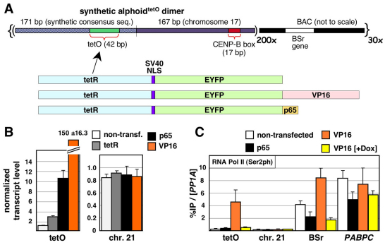
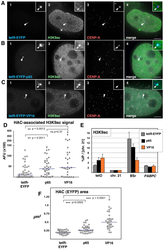
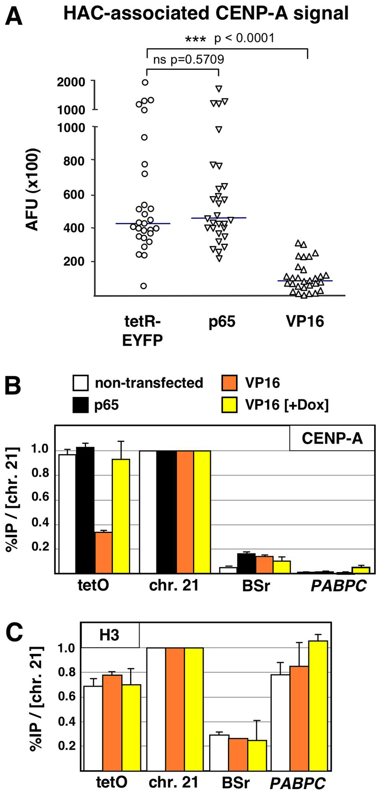
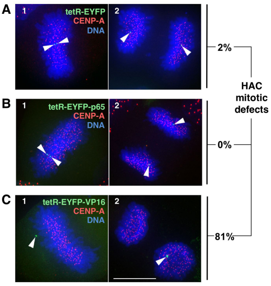
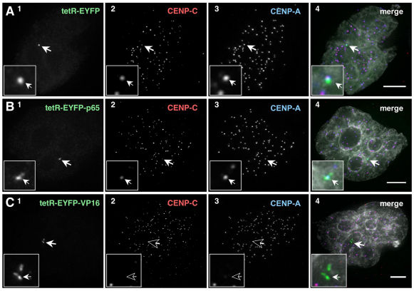
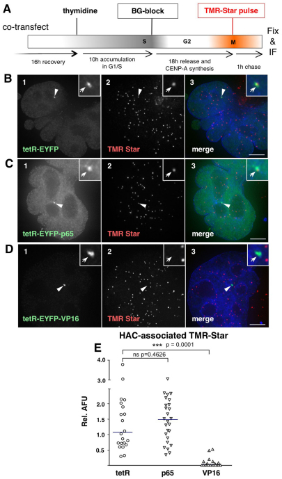
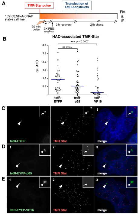
Human kinetochores are transcriptionally active, producing very low levels of transcripts of the underlying alpha-satellite DNA. However, it is not known whether kinetochores can tolerate acetylated chromatin and the levels of transcription that are characteristic of housekeeping genes, or whether kinetochore-associated 'centrochromatin', despite being transcribed at a low level, is essentially a form of repressive chromatin. Here, we have engineered two types of acetylated chromatin within the centromere of a synthetic human artificial chromosome. Tethering a minimal NF-κB p65 activation domain within kinetochore-associated chromatin produced chromatin with high levels of histone H3 acetylated on lysine 9 (H3K9ac) and an ~10-fold elevation in transcript levels, but had no substantial effect on kinetochore assembly or function. By contrast, tethering the herpes virus VP16 activation domain produced similar modifications in the chromatin but resulted in an ~150-fold elevation in transcripts, approaching the level of transcription of an endogenous housekeeping gene. This rapidly inactivated kinetochores, causing a loss of assembled CENP-A and blocking further CENP-A assembly. Our data reveal that functional centromeres in vivo show a remarkable plasticity--kinetochores tolerate profound changes to their chromatin environment, but appear to be critically sensitive to the level of centromeric transcription.


Figures

Fig. 1.
Fig. 1.
Tethering of p65 and VP16 into the alphoidtetO HAC kinetochore creates two distinct types of ‘open’ chromatin. (A) Schematic drawings of the alphoidtetO DNA array and tetR fusion constructs expressed in HeLa 1C7 cells. The synthetic monomer was based on a published consensus sequence for alphoid DNA (Choo et al., 1991). (B) Real-time RT-PCR analysis of non-transfected 1C7 cells stably carrying the alphoidtetO HAC and 2 days after transfecting constructs expressing tetR-EYFP (tetR), tetR–EYFP–p65 (p65) or tetR–EYFP–VP16 (VP16). We selected for transfected cells expressing the tethered p65 and VP16 constructs using an IRES-Puro vector by puromycin selection. Expression levels of the alphoidtetO array (tetO) and the chromosome 21 centromere (chr. 21) were normalized to those of β-actin. AlphoidtetO RNA levels in untransfected cells were arbitrarily set as 1.0. Data represent the mean and s.e.m. of three or more independent experiments. (C) ChIP analysis of nontransfected 1C7 cells or cells transfected as in B using an antibody against the elongating form of RNA polymerase II. Transfections with tetR–EYFP–VP16 were performed either in the absence of or in the presence (+Dox) of 1 μg/ml doxycycline, which prevents the binding of tetR to the alphoidtetO array. RNA polymerase II association with alphoidtetO (tetO), chromosome 21 alphoid (chr. 21) DNA, the blasticidin S resistance (BSr) marker in the BAC vector and the endogenous PABPC1 gene was determined. Values were normalized to the RNA polymerase II occupancy at the endogenous PP1A housekeeping gene locus. Data represent the mean and s.e.m. of two or more independent experiments.
Fig. 2.
Fig. 2.
Tethering of VP16 and p65 mediates HAC centromere hyper-acetylation. (A–C) Immunofluorescence (IF) analysis of 1C7 cells 2 days after transfection with the indicated tetR fusion constructs. Cells were co-stained with antibodies against acetylated H3K9 (H3K9ac, green, panel 2) and CENP-A (red, panel 3). Arrowheads depict the HAC, as determined by the EYFP signal (blue, panel 1). Merged images (panel 4) represent the overlay of EYFP signals with antibody and 4′,6-diamidino-2-phenylindole (DAPI) staining (light grey). Scale bars: 5 μm. (D) Fluorescence signals of HAC-associated H3K9ac staining in individual cells transfected as in A–C were quantified and plotted as arbitrary fluorescence units (AFU). Solid lines indicate the median. (E) ChIP analysis as in Fig. 1C of 1C7 cells transfected with tetR–EYFP, tetR–EYFP–p65 (p65) or tetR–EYFP–VP16 (VP16), using an antibody against H3K9ac. Values are normalized to the chromosome 21 centromere locus and represent the mean and s.e.m. of three independent experiments. (F) Quantification of HAC decondensation in cells transfected as in A–C. The HAC-bound EYFP area was measured in maximum-intensity projections of cells expressing the indicated fusion constructs. Sold lines indicate the median.
Fig. 3.
Fig. 3.
Tethering of VP16, but not p65, causes a specific loss of CENP-A from the alphoidtetO HAC centromere. (A) 1C7 cells were transfected with the indicated tetR fusion constructs and fixed after 2 days. CENP-A immunofluorescence staining associated with the HAC was quantified. Solid lines indicate the median. (B,C) ChIP analysis as in Fig. 1C, using antibodies against CENP-A (B) or canonical histone H3 (C). ns, no significant difference.
Fig. 4.
Fig. 4.
Tethering of p65 is compatible with HAC kinetochore function. (A–C) Cells expressing the indicated tetR fusion proteins were fixed and stained 2 days after transfection. HAC sister chromatids (arrowheads) were analyzed in metaphase (panel 1) and late anaphase or telophase (panel 2). The percentages of HACs with mitotic defects, determined as unaligned HACs in metaphase cells (e.g. C1) or mis-segregated HAC sister chromatids (e.g. C2) are indicated (n=53, 44 and 42 for tetR–EYFP, tetR–EYFP–p65 and tetR–EYFP–VP16, respectively). Scale bar: 5 μm.
Fig. 5.
Fig. 5.
Tethered VP16, but not p65, removes CENP-C from the HAC kinetochore. (A–C) Cells expressing the indicated tetR fusion proteins were fixed and stained 2 days after transfection. tetR–EYFP fusion protein (green, panel 1), CENP-C (red, panel 2) and CENP-A (blue, panel 3). Merged images (panel 4) represent the overlay of EYFP signal, antibody and DAPI staining (light grey). Arrows depict the HAC. Scale bars: 5 μm.
Fig. 6.
Fig. 6.
VP16, but not p65 tethering, negatively affects CENP-A loading at the HAC centromere. (A) Schematic diagram of quench–pulse-chase experiments in cells co-transfected with SNAP-tagged CENP-A and either of the tetR fusion constructs to determine the centromeric loading of newly synthesized CENP-A. (B–D) Cells expressing the indicated tetR fusion construct (green, panel 1) and pulse-labelled with TMR-Star (red, panel 2). Merged images (panel 3) represent the overlay of EYFP signal with TMR and DAPI staining (blue). Arrowheads depict the HAC. Scale bar: 5 μm. (E) Quantification of HAC-associated TMR-Star signal in cells as in B–D. AFU values are plotted relative to the average TMR-Star signal measured at endogenous centromeres. Solid lines indicate the median. ns, no significant difference.
Fig. 7.
Fig. 7.
VP16 causes an increased rate of loss of CENP-A from the HAC centromere. (A) Schematic diagram of pulse-chase experiments in cells stably expressing SNAP-tagged CENP-A and transfected with the various tetR fusion constructs. (B) Quantification of HAC-associated TMR-Star signal in cells plotted relative to the average TMR-Star signal measured at ten endogenous centromeres. Solid lines indicate the median. (C–E) Cells expressing the indicated tetR fusion construct (green, panel 1) and pulse-labelled with TMR-Star (red, panel 2). Merged images (panel 3) represent the overlay of EYFP signal with TMR Star and DAPI staining (blue). Arrowheads depict the HAC. Scale bar: 5 μm.

Similar articles

Cited by

References

    1. Allshire R. C., Karpen G. H. (2008). Epigenetic regulation of centromeric chromatin: old dogs, new tricks? Nat. Rev. Genet. 9, 923-937 - PMC - PubMed
    1. Alonso A., Fritz B., Hasson D., Abrusan G., Cheung F., Yoda K., Radlwimmer B., Ladurner A. G., Warburton P. E. (2007). Co-localization of CENP-C and CENP-H to discontinuous domains of CENP-A chromatin at human neocentromeres. Genome Biol. 8, R148 - PMC - PubMed
    1. Bergmann J. H., Rodriguez M. G., Martins N. M., Kimura H., Kelly D. A., Masumoto H., Larionov V., Jansen L. E., Earnshaw W. C. (2011). Epigenetic engineering shows H3K4me2 is required for HJURP targeting and CENP-A assembly on a synthetic human kinetochore. EMBO J. 30, 328-340 - PMC - PubMed
    1. Black B. E., Bassett E. A. (2008). The histone variant CENP-A and centromere specification. Curr. Opin. Cell Biol. 20, 91-100 - PubMed
    1. Black B. E., Foltz D. R., Chakravarthy S., Luger K., Woods V. L., Jr, Cleveland D. W. (2004). Structural determinants for generating centromeric chromatin. Nature 430, 578-882 - PubMed

Publication types

MeSH terms