. 2012 Apr;32(8):1442-52.
doi: 10.1128/MCB.06323-11. Epub 2012 Feb 13.

MacroH2A1 regulates the balance between self-renewal and differentiation commitment in embryonic and adult stem cells

Affiliations

MacroH2A1 regulates the balance between self-renewal and differentiation commitment in embryonic and adult stem cells

Catherine Creppe et al. Mol Cell Biol. 2012 Apr.

Abstract

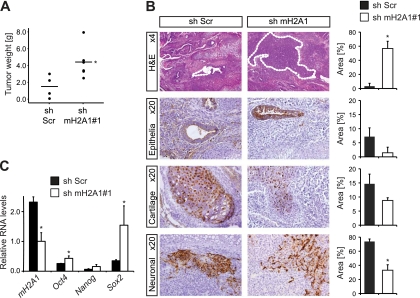
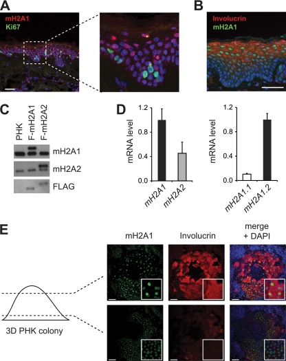
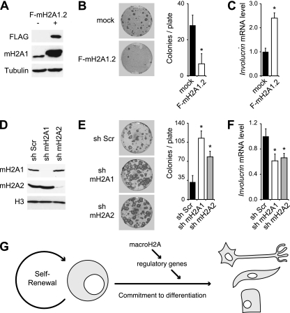
One of the most striking epigenetic alterations that occurs at the level of the nucleosome is the complete exchange of the canonical H2A histones for the macroH2A variant. Here, we provide insight into the poorly recognized function of macroH2A in transcriptional activation and demonstrate its relevance in embryonic and adult stem cells. Knockdown of macroH2A1 in mouse embryonic stem (mES) cells limited their capacity to differentiate but not their self-renewal. The loss of macroH2A1 interfered with the proper activation of differentiation genes, most of which are direct target genes of macroH2A. Additionally, macroH2A1-deficient mES cells displayed incomplete inactivation of pluripotency genes and formed defective embryoid bodies. In vivo, macroH2A1-deficient teratomas contained a massive expansion of malignant, undifferentiated carcinoma tissue. In the heterogeneous culture of primary human keratinocytes, macroH2A1 levels negatively correlated with the self-renewal capacity of the pluripotent compartment. Together these results establish macroH2A1 as a critical chromatin component that regulates the delicate balance between self-renewal and differentiation of embryonic and adult stem cells.


Figures

Fig 1
Fig 1
MacroH2A1.2 is enriched on genes that regulate differentiation in ES cells. (A) Lysates from E14 mouse embryonic stem (mES) cells were analyzed by Western blotting together with extracts containing similar amounts of Flag (F)-epitope-tagged macroH2A1.2 and macroH2A2 (marked with arrowheads). (B) The relative mRNA levels of macroH2A forms were analyzed by quantitative RT-PCR and normalized to two housekeeping genes and equimolar reference samples. (C) MspI restriction fragment length polymorphism analysis of an RT-PCR product spanning the variant exon allows the macroH2A1 splice forms to be distinguished. Murine cells expressing exclusively macroH2A1.1 were included as control. (D) Enriched regions (peaks) of macroH2A1 identified by massive parallel sequencing of DNA immunoprecipitated by anti-macroH2A1 from chromatin are shown in respect to annotated genes. (E) The positions of the peaks shown in panel D were plotted against annotated transcription start sites (TSS). (F) Correlation of mH2A1 occupancy with the transcriptional level. Genes were grouped into five expression categories according to probe intensities on Agilent Expression arrays (0, no signal/expression; 1 to 4, very low to high expression). (G) DAVID functional annotation cluster analysis of macroH2A1 target genes. All clusters with an enrichment score of >2 are represented. Asterisks indicate the P value of the top-ranked component of each cluster: *, P < 10−4; **, P < 10−5; and ***, P < 10−6. Gene examples are given at the right. For more details on panels D to G, see File S1 in the supplemental material.
Fig 2
Fig 2
MacroH2A is retained on its target genes upon induction of neuronal differentiation. Cells were treated with 1 μM retinoic acid (RA) for 3 days. (A) Western blot analysis of macroH2A1 and macroH2A2 in total nuclei. Oct4 was included as a differentiation control, and histone H3 was used as a loading control. (B) The relative mRNA levels of total macroH2A1, macroH2A2, and the splice variants macroH2A1.1 and macroH2A1.2 were analyzed by quantitative RT-PCR and normalized to two housekeeping genes and equimolar reference sampled. (C) The occupancy of macroH2A1 on genes was analyzed by chromatin immunoprecipitation (ChIP) in self-renewing cells and differentiating cells treated with 1 μM RA for 3 days. Error bars denote standard deviation, and n = 3. (D) The mRNA level of Pax3 and the occupancy by macroH2A1 around the transcription start site of Pax3 were analyzed by quantitative RT-PCR and ChIP, respectively. Error bars denote standard deviation, and n = 3.
Fig 3
Fig 3
Depletion of macroH2A1 interferes with the neuronal differentiation of mES cells. (A) Expression levels of differentiation and pluripotency genes were analyzed by RT-qPCR in mES cells that were untreated or treated with 1 μM RA for 3 days. Upp1, Srcap, and Hmg20a were included as controls. Error bars denote standard deviation; n = 3; *, P < 0.05. (B) Crude cell lysates were analyzed by Western blotting using anti-Oct4, macroH2A1, and histone H3 antibodies. (C) mES cells were treated with 1 μM RA for 6 days, after which RA medium was removed and replaced by LIF maintenance mES cell medium. Cells were collected and subjected to RNA extraction and quantitative RT-PCR (17). Error bars denote the variances of results from two independent experiments.
Fig 4
Fig 4
Depletion of macroH2A1 interferes with proper embryoid body formation. (A) Western blot analysis of macroH2A1 and Oct4 in self-renewing mES cells and in embryoid bodies (EBs) at day 9. Histone H3 was used as loading control. (B) The mRNA levels of macroH2A1 and macroH2A2 in control cells and after shRNA-mediated knockdown of macroH2A1 were analyzed at different time points of EB formation and normalized to equimolar reference samples. Error bars denote standard deviation; n = 3; *, P < 0.05. (C) Light microscopic analysis of EBs revealed that the normal shapes and sizes are perturbed in macroH2A1-deficient cells. Scale bar, 500 μm. Arrowheads indicate normal stage-specific cavitation. (D) Transcript expression levels of genes that regulate pluripotency (Nanog, Oct4, and Sox2) and differentiation (T/Brachyury, mesoderm; Gata4, endoderm; Hand1, Pax3, and nestin, ectoderm; and Cdx2, trophoectoderm) were analyzed by quantitative RT-PCR. Error bars denote standard deviation; n = 3; *, P < 0.05. (E) The mRNA levels of Nanog, Gata4, macroH2A1, and macroH2A2 were analyzed in ES cells expressing different hairpins directed against macroH2A1 and macroH2A2 at day 6 of EB formation (day 9 for Nanog). Error bars denote standard deviation; n = 3; *, P < 0.05.
Fig 5
Fig 5
In vivo loss of macroH2A1 results in an expansion of malignant, undifferentiated carcinoma tissue. (A) Weight of teratomas generated by sh-macroH2A1 and control mES cells 3 weeks after injection.*, P = 0.019, as determined by nonparametric Mann-Whitney testing. (B) Hematoxylin-eosin (H&E) stainings and immunohistochemical analyses of teratoma sections. Malignant undifferentiated carcinoma tissues were identified by two independent pathologists and indicated by dotted lines. Different markers were used to stain tissues originating from all three lineages: S100 for cartilage (mesoderm), glial fibrilary acidic protein for neuroglia (ectoderm), and cytokeratins for epithelial cells (endoderm). Quantifications on the right were done on complete cross-sections of three samples. Error bars denote standard deviation; *, P < 0.05. (C) Quantitative RT-PCR analysis of RNA isolated from bulk teratoma tissue. Values are shown with respect to the mRNA levels in self-renewing stem cells, which were set to 1. Error bars denote standard deviation; n = 3; *, P < 0.05.
Fig 6
Fig 6
MacroH2A1 expression correlates with the differentiation of human keratinocytes. (A and B) Gradual upregulation of macroH2A1 from the basal to the more differentiated cell layers in a human scalp skin section, as shown by immunofluorescence. Cells were counterstained for the proliferation marker Ki67 (A) or the differentiation marker involucrin (B) and DAPI. Scale bars, 50 μm. (C) Lysates from primary human keratinocytes (PHKs) were analyzed by Western blotting together with extracts containing equivalent amounts of Flag (F)-tagged macroH2A1.2 and macroH2A2. (D) The relative mRNA levels of macroH2A forms were analyzed by quantitative RT-PCR and normalized to equimolar reference samples. (E) Confocal microscopy sections of a three-dimensional PHK colony. Involucrin was used to mark the differentiated cells present in the upper layer. DNA was counterstained with DAPI. Scale bars, 100 μm.
Fig 7
Fig 7
MacroH2A1 limits the self-renewal capacity, and favors the differentiation, of primary human keratinocytes. (A) Western blot analysis of lysates from PHKs overexpressing Flag (F)-tagged macroH2A1.2 using anti-FLAG, macroH2A1, and tubulin antibodies. (B) Colony formation analysis of the same PHKs grown on feeder cells. Error bars denote standard deviation; n = 3; *, P < 0.05. (C) The mRNA level of involucrin was analyzed by quantitative RT-PCR. Error bars denote standard deviation; n = 3; *, P < 0.05. (D) Efficient knockdown of macroH2A1 and macroH2A2 was assessed by Western blot. (E) Colony formation analysis of the same cells. Error bars denote standard deviation; n = 3; *, P < 0.05. (F) Expression analysis of involucrin by quantitative RT-PCR. Error bars denote standard deviation; n = 3; *, P < 0.05. (G) Cartoon illustrating how macroH2A affects the balance of self-renewal and differentiation. The presence of macroH2A on regulatory differentiation facilitates their activation and thereby the commitment of stem cells to differentiation.

References

    1. Agelopoulos M, Thanos D. 2006. Epigenetic determination of a cell-specific gene expression program by ATF-2 and the histone variant macroH2A. EMBO J. 25:4843–4853 - PMC - PubMed
    1. Barde I, Salmon P, Trono D. 2010. Production and titration of lentiviral vectors. Curr. Protoc. Neurosci. 53:4.21.1–4.21.23. - PubMed
    1. Barrandon Y, Green H. 1987. Three clonal types of keratinocyte with different capacities for multiplication. Proc. Natl. Acad. Sci. U. S. A. 84:2302–2306 - PMC - PubMed
    1. Blum B, Bar-Nur O, Golan-Lev T, Benvenisty N. 2009. The anti-apoptotic gene survivin contributes to teratoma formation by human embryonic stem cells. Nat. Biotechnol. 27:281–287 - PubMed
    1. Boix R, et al. 2009. Primary renal cell carcinoma in a transplanted kidney: genetic evidence of recipient origin. Transplantation 87:1057–1061 - PubMed

Publication types

MeSH terms