

doi: 10.1128/MCB.05641-11.
Epub 2012 Feb 13.
PinX1 localizes to telomeres and stabilizes TRF1 at mitosis
Affiliations
- PMID: 22331467
- PMCID: PMC3318592
- DOI: 10.1128/MCB.05641-11
Item in Clipboard
PinX1 localizes to telomeres and stabilizes TRF1 at mitosis
Mol Cell Biol.
2012 Apr.
Abstract
Human telomeres are DNA-protein complexes that cap and protect the ends of chromosomes. The protein PinX1 associates with telomeres through an interaction with the resident double-stranded telomere-binding protein TRF1. PinX1 also binds to and inhibits telomerase, the enzyme responsible for complete replication of telomeric DNA. We now report that endogenous PinX1 associates with telomeres primarily at mitosis. Moreover, knockdown of PinX1 caused delayed mitotic entry and reduced the accumulation of TRF1 on telomeres during this stage of the cell cycle. Taking these findings together, we suggest that one function of PinX1 is to stabilize TRF1 during mitosis, perhaps to promote transition into M phase of the cell cycle.
Figures
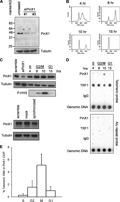
PinX1 localizes at telomeres during M phase. (A) Levels of endogenous PinX1, as assessed by immunoblot assay with an anti-PinX1 antibody, in cells untreated or treated with two different PinX1 siRNAs. Data are representative of two replicate experiments. (B to D) HeLa cells were synchronized by double thymidine block, released, and collected at the four indicated time points. Collected cells were split into three portions that were stained with propidium iodide and subjected to FACS for cell cycle analysis (B), immunoblotted to detect endogenous PinX1, phospho-histone H3 (P-HH3) as a marker of mitosis, or tubulin as a loading control (as additional controls, PinX1 and tubulin levels were also analyzed in scrambled-siRNA-transfected cells, mock-transfected cells, or untransfected cells synchronized by double thymidine block) (C), or used for ChIP analysis with an anti-PinX1 antibody or an anti-TRF1 antibody or IgG as controls, followed by Southern hybridization with a telomere or control Alu probe (5 μg genomic DNA served as a hybridization control) (D). Data are representative of three replicate experiments. (E) Means ± SD of the normalized hybridization intensities of telomeric DNA coimmunoprecipitated with endogenous PinX1 from three independent ChIP experiments at the indicated cell cycle phases.
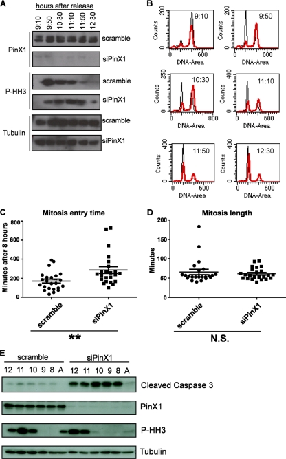
Knockdown of PinX1 delays entry into mitosis. HeLa cells transfected with a scrambled control siRNA or PinX1 siRNA were synchronized by double thymidine block, released, and collected at the indicated six time points. (A and B) Lysates from cells at each time point and asynchronous cells were either immunoblotted to detect endogenous PinX1, phosphorylated histone H3 (P-HH3) as a marker of mitosis, or tubulin as a loading control (A) or stained with propidium iodide and subjected to FACS for cell cycle analysis (black trace, scrambled-siRNA-treated control cells; red trace, PinX1 siRNA-treated cells) (B). Data are representative of five replicate experiments. (C and D) Mean length of time ± SD to enter (C) or to complete (D) mitosis beginning 8 h after release from a double thymidine block for 23 HeLa cells stably expressing GFP-histone H2B and transfected (as verified by BLOCK-iT Alexa Fluor Red oligonucleotide fluorescence) with either a scrambled control siRNA (●) or a PinX1 siRNA (■). **, P < 0.01; N.S., not significant. (E) HeLa cells transfected with a scrambled control siRNA or a PinX1 siRNA were synchronized by double thymidine block, released, collected at the time points indicated above the gels, and immunoblotted to detect endogenous PinX1, phosphorylated histone H3 (P-HH3), and cleaved caspase 3. Data are representative of two replicate experiments. A, asynchronous cells.
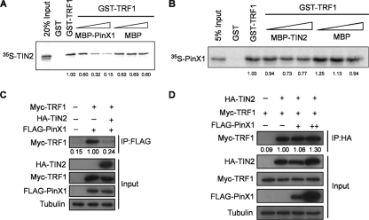
TIN2 inhibits PinX1 binding to TRF1. (A) Association of rabbit reticulocyte lysate (RRL)-generated 35S-labeled recombinant TIN2 with bacterially generated recombinant GST-TRF1 upon coincubation with increasing concentrations of recombinant MBP-PinX1. GST and MBP served as controls. The relative amounts of TIN2 normalized to the amount detected with GST-TRF1 are shown below the gel. Data are representative of two replicate experiments. (B) Association of RRL-generated 35S-labeled recombinant PinX1 with bacterially generated recombinant GST-TRF1 upon coincubation with increasing concentrations of recombinant MBP-TIN2. GST and MBP served as controls. The relative amounts of PinX1 normalized to the amount detected with GST-TRF1 are shown below the gel. Data are representative of two replicate experiments. (C) Levels of Myc-TRF1 detected by immunoprecipitation (IP) of FLAG-PinX1 followed by immunoblot assay with an anti-Myc antibody in 293T cells expressing HA-TIN2. Tubulin served as a loading control. The relative amounts of Myc-TRF1 coimmunoprecipitated with FLAG-PinX1 normalized to tubulin are shown below the gel. Data are representative of two replicate experiments. (D) Levels of Myc-TRF1 detected by immunoprecipitation of HA-TIN2 followed by immunoblot assay with an anti-Myc antibody in 293T cells expressing FLAG-PinX1. Tubulin served as a loading control. The relative amounts of Myc-TRF1 coimmunoprecipitating with HA-TIN2 normalized to tubulin are shown below the gel. Data are representative of two replicate experiments.
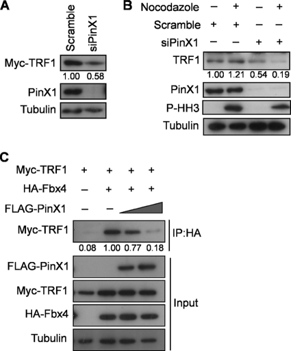
Knockdown of PinX1 decreases TRF1 levels at M phase. (A) Myc-TRF1 levels in HeLa cells treated with PinX1 or scramble siRNA, as assessed by immunoblot assay. Tubulin served as a loading control. The relative amounts of Myc-TRF1 normalized to tubulin are shown below the gel. Data are representative of two replicate experiments. (B) HeLa cells treated with a scrambled control siRNA or PinX1 siRNA and either left untreated (interphase cells) or treated with nocodazole and subjected to mitotic shake-off (mitotic cells) were immunoblotted to detect phospho-histone H3 (P-HH3) as a mitotic marker, PinX1, TRF1, or tubulin as a loading control. The relative amounts of TRF1 normalized to tubulin are shown below the gel. Data are representative of two replicate experiments. (C) Amount of Myc-TRF detected by immunoprecipitation (IP) of HA-Fbx4 followed by immunoblot assay with an anti-Myc antibody in 293T cells with increasing amounts of FLAG-PinX1. Tubulin served as a loading control. The relative amounts of Myc-TRF1 coimmunoprecipitated with HA-Fbx4 normalized to tubulin are shown below the gel. Data are representative of three replicate experiments.
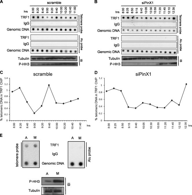
Knockdown of PinX1 delays the reloading of TRF1 onto telomeres during mitosis. HeLa cells transfected with a scrambled control siRNA (A) or a PinX1 siRNA (B) were synchronized by double thymidine block, released, and collected at the indicated time points after release from the block. Collected cells were split into two portions for ChIP and immunoblot analysis. ChIP analysis was performed with an anti-TRF1 antibody or IgG as a control, followed by Southern hybridization with a telomere or control Alu probe (genomic DNA served as a hybridization control). Immunoblot analysis was performed to detect phosphorylated histone H3 (P-HH3) as a marker of mitosis or tubulin as a loading control. Data are representative of five (A) and three (B) replicate experiments. (C and D) Normalized hybridization intensities of telomeric DNA coimmunoprecipitating with endogenous PinX1 at the indicated time points in scrambled (C) or PinX1 (D) siRNA-treated cells. (E) Asynchronous (interphase) HeLa cells or HeLa cells treated with nocodazole and subjected to mitotic shake-off to enrich metaphase (M) cells were subjected to ChIP analysis and immunoblotted for phosphorylated histone H3 or tubulin as described above. Data are representative of two replicate experiments.
Similar articles
-
Telomerase inhibitor PinX1 provides a link between TRF1 and telomerase to prevent telomere elongation.J Biol Chem. 2011 Feb 4;286(5):3894-906. doi: 10.1074/jbc.M110.180174. Epub 2010 Nov 30. J Biol Chem. 2011. PMID: 21119197 Free PMC article.
-
PinX1, a telomere repeat-binding factor 1 (TRF1)-interacting protein, maintains telomere integrity by modulating TRF1 homeostasis, the process in which human telomerase reverse Transcriptase (hTERT) plays dual roles.J Biol Chem. 2014 Mar 7;289(10):6886-6898. doi: 10.1074/jbc.M113.506006. Epub 2014 Jan 10. J Biol Chem. 2014. PMID: 24415760 Free PMC article.
-
Human PinX1 mediates TRF1 accumulation in nucleolus and enhances TRF1 binding to telomeres.J Mol Biol. 2009 May 22;388(5):928-40. doi: 10.1016/j.jmb.2009.02.051. Epub 2009 Mar 3. J Mol Biol. 2009. PMID: 19265708
-
Role of Pin2/TRF1 in telomere maintenance and cell cycle control.J Cell Biochem. 2003 May 1;89(1):19-37. doi: 10.1002/jcb.10496. J Cell Biochem. 2003. PMID: 12682905 Review.
-
Post-translational modifications of TRF1 and TRF2 and their roles in telomere maintenance.Mech Ageing Dev. 2012 Jun;133(6):421-34. doi: 10.1016/j.mad.2012.05.002. Epub 2012 May 23. Mech Ageing Dev. 2012. PMID: 22634377 Review.
Cited by
-
Prognostic and Clinicopathological Value of PINX1 in Various Human Tumors: A Meta-Analysis.Biomed Res Int. 2018 Jul 16;2018:4621015. doi: 10.1155/2018/4621015. eCollection 2018. Biomed Res Int. 2018. PMID: 30079348 Free PMC article.
-
The truncated isoform of the receptor for hyaluronan-mediated motility (RHAMMΔ163) modulates shelterin and telomerase reverse transcriptase transcription affecting telomerase activity.Front Aging. 2025 Jun 30;6:1604051. doi: 10.3389/fragi.2025.1604051. eCollection 2025. Front Aging. 2025. PMID: 40661163 Free PMC article.
-
The 68-kDa telomeric repeat binding factor 1 (TRF1)-associated protein (TAP68) interacts with and recruits TRF1 to the spindle pole during mitosis.J Biol Chem. 2014 May 16;289(20):14145-56. doi: 10.1074/jbc.M113.526244. Epub 2014 Apr 1. J Biol Chem. 2014. PMID: 24692559 Free PMC article.
-
Integrating data and knowledge to identify functional modules of genes: a multilayer approach.BMC Bioinformatics. 2019 May 2;20(1):225. doi: 10.1186/s12859-019-2800-y. BMC Bioinformatics. 2019. PMID: 31046665 Free PMC article.
-
Cell cycle regulated phosphorylation of the telomere-associated protein TIN2.PLoS One. 2013 Aug 16;8(8):e71697. doi: 10.1371/journal.pone.0071697. eCollection 2013. PLoS One. 2013. PMID: 23977114 Free PMC article.
References
-
- Banik SS, Counter CM. 2004. Characterization of interactions between PinX1 and human telomerase subunits hTERT and hTR. J. Biol. Chem. 279: 51745–51748 - PubMed
Publication types
MeSH terms
Substances
Grants and funding
LinkOut - more resources
Full Text Sources
Miscellaneous